Atlas of Rashes Associated With Fever (Pictures)
Last reviewed by Dr. Raj MD on January 12th, 2022.
This atlas is an attempt to depict the most common and important rashes that we see in our daily life. It necessarily is not in order of importance.
Neither is it possible to describe all the rashes which we see. Nevertheless, we have given an honest effort to collate the most important ones here.

Streptococcal Toxic Shock Syndrome
Toxic shock syndrome, as the name indicates, is toxin mediated. Organisms such as Group A Streptococcus (Streptococcus pyogenes) and Staphylococcus aureus can cause this condition. Streptococcus pyogenes produce a toxin Streptococcal pyrogenic exotoxin.
The toxin when passed into the circulation results in hypotension, high fever, multi organ failure (3 or more organ systems are involved), vomiting, diarrhoea, headache and rashes. There can also be skin peeling out of palms and soles [1].
Streptococcal Toxic Shock Syndrome Rash Pictures
Photo 1: Toxic shock syndrome rash on leg.
Image Source: syndromespedia.com
Image 2: Toxic shock syndrome rash on the trunk of a patient.
Photo Source: syndromespedia.com
Picture 3: Desquamation in palm due to toxic shock syndrome.
Image Source: 2.bp.blogspot.com
Scarlet fever (Second Disease/ Scarlatina)
The disease is caused by Streptococcus pyogenes. The toxins produced by this bacterium can cause Scarlet fever. It usually follows a throat infection by the same bacteria. Sore throat with swollen glands, high fever, rashes, flushed face, pallor around the mouth and strawberry like tongue could be scarlet fever.
A doctor has to confirm the diagnosis after appropriate examination and investigations. The rashes generally start from the face or neck and then spread down to the limbs and trunk[2].
Scarlet fever (Second Disease/ Scarlatina) Rash Pictures
Image 4: Rashes of scarlet fever over neck and trunk.
Picture Source: kidspot.co.nz
Photo 5: ‘Strawberry tongue’ in a child with Scarlet fever
Image Source: tonguepictures.org
Picture 6: Scarlet fever rashes on upper limbs and back
Photo Source: : www.drugs.com
Image 7: Scarlet fever rash on a child’s face.
Picture Source: ichef-1.bbci.co.uk
Staphylococcal Toxic Shock Syndrome
Here, the organism causing toxic shock syndrome is Staphylococcus aureus. The toxin produced by staphylococcus which causes this condition is Toxic Shock Syndrome Toxin-1 (TSST-1). The toxin when passed into the circulation results in hypotension, high fever, multi organ failure (3 or more organ systems are involved), vomiting, diarrhoea, headache and rashes.
There can also be skin peeling out of palms and soles[1].
Staphylococcal Toxic Shock Syndrome Rash Pictures
Photo 8: Desquamation (skin peeling off) of palm can be seen in a case of Staphylococcal toxic shock syndrome.
Image Source: www.nejm.org
Image 9: Toxic shock syndrome rashes on abdomen.
Picture Source: www.ladycarehealth.com
Picture 10: Desquamation (skin peeling off) of palm can be seen in a case of Staphylococcal toxic shock syndrome.
Photo Source: www.primehealthchannel.com
Image 11: Rashes in Toxic shock syndrome.
Picture Source: 4.bp.blogspot.com
Staphylococcal Scalded Skin Syndrome
The disease is caused due to a toxin, Epidermolytic toxin produced by Staphylococcus aureus. The toxin spreads through the blood and can cause lesions that resemble scalding (burns caused by hot water or liquids). Mostly it is seen in children below 5 years of age though it can affect any aged person. In new born, the condition is also called as Ritter’s disease.
The child may present with fever, irritability, redness of the skin and fluid filled blisters. Peeling off of large areas of skin may occur [3].
Staphylococcal Scalded Skin Syndrome Rash Pictures
Photo 12: Staphylococcal scalded skin syndrome
Image Source: www.researchgate.net
Image 13: Staphylococcal scalded skin syndrome
Picture Source: www.nejm.org
Photo 14: Staphylococcal scalded skin syndrome
Image Source: www.alpfmedical.info
Picture 15: Staphylococcal scalded skin syndrome
Photo Source: cmr.asm.org
Pseudomonas “Hot-Tub” Folliculitis
‘Follicle’ is the lower part of a hair strand. When an infection occurs in the follicle, it is called as folliculitis. In people frequently using a hot tub or swimming pool, Pseudomonas aeruginosa can get to the base of hair and cause folliculitis.
It can be seen as vesicles or pustules at the follicles of areas which come in contact with water for a longer time. Generally it involves abdomen, buttocks, axillae etc[4].
Pseudomonas “Hot-Tub” Folliculitis Rash Pictures
Photo 16: Hot Tub Folliculitis
Image Source: www.clean-pool-and-spa.com
Picture 17: Hot Tub folliculitis
Photo Source: www.clean-pool-and-spa.com
Image 18: Hot Tub Folliculitis. Note the vesicle at the root of hair follicles
Picture Source: www.wrinkle-free-skin-tips.com
Photo 19: Hot tub folliculitis
Image Source: www.huidziekten.nl
Ecthyma gangrenosum
Ecthyma Gangrenosum, most commonly caused by Pseudomonas aeruginosa, is usually seen in patients with low immunity. When the immunity is low any infection can easily occur. The organism enters through a break in skin or through infection in any other parts of the body.
The lesion seen is initially a macule (<1 cm wide skin discoloration) which rapidly aggravates into a haemorrhagic pustule. The area degenerates (gangrene formation) with surrounding erythema (redness) and central scar (eschar) formation[5].
Ecthyma gangrenosum Rash Pictures
Image 20: Ecthyma gangrenosum. Eschar can be seen in the picture.
Picture Source: www.atlasdermatologico.com
Photo 21: Ecthyma gangrenosum. Eschar can be seen in the picture.
Image Source: www.atlasdermatologico.com
Picture 22: Ecthyma gangrenosum.
Photo Source: mddk.com
Image 23: Ecthyma gangrenosum.
Picture Source: mddk.com
Thrombotic thrombocytopenic purpura (TTP)
The three parts of the name make it easy to understand the condition. Due to thrombosis (blockage of small blood vessels), there is a decrease in platelet count. When platelet count decreases, there can be minute bleeding into the layers of skin called as petechiae.
Along with petechiae, there can be central nervous system involvement (seizures, paraesthesia, visual disturbances etc) and anaemia (fatigue)[6, 7].
Thrombotic thrombocytopenic purpura (TTP) Rash Pictures
Photo 24: Thrombotic thrombocytopenic purpura in a pregnant lady.
Image Source: : www.researchgate.net
Image 25: https://www.nhlbi.nih.gov/sites/www.nhlbi.nih.gov/files/images_273
Thrombotic thrombocytopenic purpura.
Picture 26: Thrombotic thrombocytopenic purpura.
Photo Source: www.thromboticthrombocytopenicpurpura.com
Tularemia
Tularemia is a zoonotic (disease spreading from animals to man) affecting rodents and hares caused by an organism Francisella tularensis. It enters human body through tick bite, ingestion of contaminated water, eyes, or by skin contact with infected animals. It is highly contagious and can be life threatening if not treated promptly.
Tularemia is characterized by lesions that are characteristic based on their route of entry. When the route of entry is skin, there can be ulceration with lymph node enlargement. Whereas, if the route of entry is eyes, then there can be irritation and inflammation of the eye. If it enters the body by ingestion of contaminated water or food, then it may cause tonsillitis, ulcerations in mouth and enlargement of the glands in neck[8, 9].
Tularemia Rash Pictures
Image 27: A case of Tularemia (oropharyngeal type) with cervical lymphadenopathy (enlarged lymph nodes)
Picture Source: www.researchgate.net
Picture 28: Tularemia under the nail due to rabbit bite.
Photo Source: img.medscapestatic.com
Anthrax
Anthrax is another zoonotic disease which is caused by an organism, Bacillus anthracis. According to the route of entry, the features of disease can be seen. On the skin, it is seen as a papule which develops into an eschar[10].
Anthrax Rash Pictures
Photo 29: Cutaneous type of anthrax with eschar on the cheek of a person.
Image Source: media.infectiousdiseaseadvisor.com
Image 30: Photo shows eschar formation in anthrax with surrounding erythema (redness)
Photo Source: www.clevelandclinicmeded.com
Picture 31: Lesion in cutaneous anthrax
Image Source: diseaseslab.com
Photo 32: Anthrax lesion
Pictue Source: tip10.info
Lyme disease
It is transmitted by ticks to humans and most commonly caused by Borrelia burgdorferi. At the site of bite, an area of redness develops and expands (Erythema migrans). Other features include joint pains, headache, palpitations, facial paralysis etc. Occasionally it may show a ‘Bull’s eye’ appearance[11].
Lyme disease Rash Pictures
Photo 33: ‘Bull’s Eye’ Appearance in Lyme’s disease.
Image Source: www.dogarmor.net
Image 34: http://cursoenarm.net
Erythema migrans seen in Lyme’s disease
Picture 35: Erythema migrans rashes
Photo Source: www.spokesman-recorder.com
Image 36: A lesion showing ‘Bull’s eye’ appearance of rash in case of Lyme’s disease.
Picture Source: media.syracuse.com
Relapsing fever
Relapsing fever is also caused by Borrelia species. Borrelia recurrentis is transmitted by louse and causes epidemic relapsing fever/louse borne relapsing fever. Whereas Borrelia hermsii and few other borrelia species is transmitted by ticks and causes endemic relapsing fever/tick borne relapsing fever.
High fever which occurs recurrently along with period of crisis due to severe hypotension marks this disease. This disease can be fatal if not treated appropriately [12, 13, 14, 15].
Relapsing fever Rash Pictures
Image 37: Rash seen in relapsing fever.
Photo Source: www.licesquad.com
Secondary syphilis
Syphilis, a sexually transmitted disease has 4 stages, primary, secondary, latent and tertiary. The secondary stage presents with rashes all over the body but mostly on palms and soles. It can also show generalized lymphadenopathy (enlarged lymph nodes) and condyloma lata (wart like lesion in the genitalia and perineum)[16, 17, 18].
Secondary syphilis Rash Pictures
Photo 38: Image shows rashes seen in secondary syphilis, in palms.
Image Source: privatetestingcenter.com
Image 39: Secondary syphilis with rash over trunk.
Photo Source: cmr.asm.org
Picture 40: Secondary syphilis- generalized rashes
Image Source: www.cmaj.ca
Image 41: Secondary syphilis- Rashes on soles.
Photo Source: www.researchgate.net
Typhoid fever
Typhoid fever is caused by Salmonella typhi and paratyphi groups which spreads generally through contaminated food or water. It presents as high fever with diarrhoea, joint pains, headache, tiredness, and anorexia. It may also show rashes called as ‘Rose spots’[19, 20].
Typhoid fever Rash Pictures
Photo 42: Rash in typhoid fever.
Image Source: quatr.us
Picture 43: Typhoid fever- Rose spots
Photo Source: onlinejindagi.files.wordpress.com
Image 44: Typhoid fever- Rose spots.
Picture Source: image.slidesharecdn.com
Rickettsial Pox
It is caused by an organism Rickettsia akari and transmitted by mites. A papule which develops into vesicles followed by ulceration and eschar formation occurs. Systemic symptoms like high fever, headache, throat pain, muscle aches, chills & rigor, vertigo, nausea etc can be seen. It usually resolves by itself [21, 22, 23].
Rickettsial Pox Rash Pictures
Photo 45: Rickettsial pox- eschar formation.
Image Source: images.emedicinehealth.com
Image 46: Rickettsial pox- eschar formation.
Picture Source: www.rugusavay.com
Photo 47: Rickettsial pox- The vesicles may resemble those in chicken pox.
Image Source: dxline.info
Rocky Mountain spotted fever
RMSF is caused by Rickettsia rickettsii. The disease usually presents with high fever, headache, muscle pain, nausea and rashes. The non-itchy petechial rashes appears on ankles and wrists which spread over to the palms, soles and other parts of the body[24, 25].
Rocky Mountain spotted fever Rash Pictures
Photo 48: Rashes of Rocky Mountain Spotted fever on a child’s forearm and hand.
Image Source: www.healthline.com
Picture 49: Petechial rashes of Rocky Mountain spotted fever on leg.
Photo Source: media.dermatologyadvisor.com
Image 50: Rocky Mountain Spotted fever- Rashes on ankles, legs and soles.
Picture Source: img.webmd.com
Typhus Fever
Epidemic typhus is a rickettsial disease caused by Rickettsia prowazekii. The organism enters the human body through louse. The symptom of the disease includes, fever, muscle pains, nausea, vomiting, lethargy and rashes. In some people, the organism may persist even after the symptoms disappear. These organisms get reactivated when the immunity decreases and can cause the recurrence called as Brill-Zinsser disease.
Endemic typhus, also known as Murine typhus, is another rickettsial disease caused by Rickettsia typhi. The organism is usually transmitted by rat flea. The symptom of the disease includes, fever, muscle pains, nausea, vomiting, lethargy and rashes[26, 27, 28, 29].
Typhus Fever Rash Pictures
Photo 51: Epidemic Typhus fever rash
Image Source: image.slidesharecdn.com
Picture 52: Epidemic Typhus fever rash
Photo Source: www.natural-health-news.com
Image 53: Epidemic Typhus fever rash
Picture Source: www.microbiologysociety.org
Picture 54: Endemic typhus
Photo Source: westjem.com
Scrub typhus
Scrub typhus is caused by Orientia tsutsugamushi. The organism enters the body through bite of a mite. An eschar formation occurs at the site of the bite. Systemic involvement including cardiac, pulmonary and central nervous system can occur. Generally a high fever, with eschar formation and constitutional symptoms such as lethargy, anorexia, nausea, headache etc occurs in an infection of scrub typhus [30].
Scrub typhus Rash Pictures
Photo 57: www.defence.gov.au
An eschar formed at the site of mite bite in case of Scrub typhus.
Photo 58: Eschar on thigh due to scrub typhus infection.
Picture Source: www.atmph.org
Image 59: Sometimes the eschar formation might be hidden even on the scalp.
Picture Source: what-when-how.com
Primary HIV infection
In individuals who are recently infected with HIV, non-specific macules and papules may be seen. This is due to skin reaction which occurs when the immunity is low. Other symptoms related to primary HIV infection may be present. Skin rashes in HIV can vary widely as it depends on the organism and cause of the condition[31, 32, 33].
Primary HIV infection Rash Pictures
Photo 60: Rash due to Kaposi’s Sarcoma in an HIV infected patient.
ImageSource: img.medscape.com
Picture 61: Herpes zoster infection in an HIV infected patient.
Photo Source: img.medscape.com
Image 62: Molluscum contagiosum in a patient with HIV
Photo Source: img.medscape.com
Photo 63: Exacerbation of psoriasis in an HIV infected patient.
Picture Source: img.medscape.com
Infectious mononucleosis
Infectious mononucleosis, also known as kissing disease, is well known for it being contagious with close contacts especially in the young. The disease is caused by Ebstein-Barr virus (Human Herpes Virus 4 /HHV4). The presentation of patient is usually with fever, sore throat, fatigue and enlarged lymph nodes. It may also show diffuse macules or papules, urticaria, petechiae and peri-orbital oedema[33, 34, 35, 36].
Infectious mononucleosis Rash Pictures
Picture 64: Infectious mononucleosis rash
Image Source: www.elsevier.es
Image 65: Rash in case of a patient with infectious mononucleosis
Photo Source: www.rocketswag.com
Picture 66: Thick white covering over enlarged tonsils, seen in case of a patient with infectious mononucleosis.
Photo Source: 5minuteconsult.com
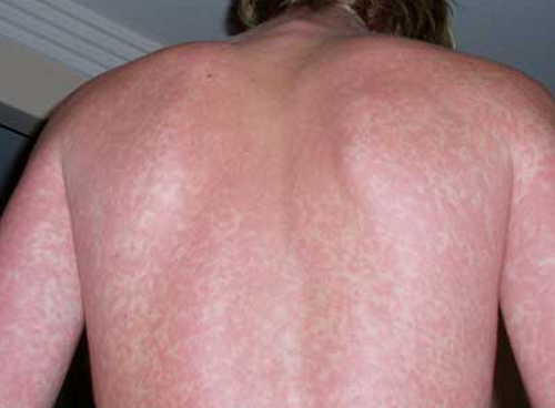
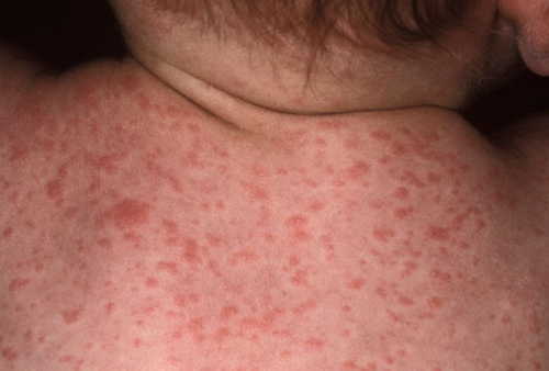
Rubeola (measles, first disease)
Measles is a highly contagious infection, caused by virus, and spreads rapidly through air (cough, sneeze, speaking etc). Around 90% of people, who come in contact with the infected person, usually get infected. The initial symptoms resemble an upper respiratory tract infection with runny nose, fever, sore throat and swelling of the lymph nodes in the neck. In 2-3 days, white spots appear in the mouth (Koplik’s spot), which are characteristic of Measles.
Rashes start to break out from the face and then spread on to neck, trunk, arm, legs and feet. Initially they are flat, sometimes may be raised from the surface, and as they spread to the other parts of the body they may become joined together[33, 37, 38, 39].
Rubeola (measles, first disease) Rash Pictures
Image 67: Measles infection in a child.
Picture Source: www.abc.net.au
Photo 68: A child showing severe measles infection
Image Source: nrvs.info
Picture 69: A closer look on the rashes due to Measles in a child.
Photo Source: tgp.com.ph
Rubella (German measles, third disease)
Rubella is also a contagious disease usually spreading among the children and the youth. It can present with rashes, lymphadenopathy (enlarged lymph nodes), fever, arthritis etc. The rashes start from the face and then spread downward. Rubella when infecting a pregnant lady can be dangerous to the child, for it may be born with cataract, heart disease and deafness[40].
Rubella (German measles, third disease) Rash Pictures
Image 70: Rubella rash
Picture Source: assets.nhs.uk
Picture 71: Rubella rash
Photo Source: images.medicinenet.com
Image 72: A closer look on the rashes due to Rubella
Picture Source: www.healthline.com
Erythema infectiosum (fifth disease)
This disease is spread through the air from infected persons. Parvovirus B19 is the organism causing this condition. Erythema infectiosum starts with fever, sore throat, runny nose, headache, abdominal pain etc. A period of around 7-10 days of no symptoms follows the initial period. Then the rashes appear on face giving the classical feature of ‘slapped cheek appearance’. Lace like reticular maculopapular rashes appear after 1-4 days[41].
Erythema infectiosum (fifth disease) Rash Pictures
Picture 73: Rash of Erythema infectiosum can be seen on the cheeks of this child.
Photo Source: www.dermquest.com
Image 74: ‘Slapped Cheek’ appearance on a baby due to Parvo B19 infection (Erythema infectiosum)
Picture Source: assets.babycenter.com
Photo 75: The rashes in Erythema infectiosum may show a lacey appearance.
Image Source: nursingfile.com
Exanthem subitum (roseola infantum, sixth disease)
Exanthem subitum is caused by Human Herpes Virus 6 (HHV6). The condition is characterized by a high fever initially which subsides abruptly within 3-5 days, followed by appearance of maculopapular rash over trunk and neck. The child may present with febrile seizures. It is usually seen in infants[42, 43, 33].
Exanthem subitum (roseola infantum, sixth disease) Rash Pictures
Image 76: Roseola infantum rash.
Photo Source: assets.nhs.uk
Photo 77: Roseola infantum
Picture Source: www.consultant360.com
Image 78: A closer look on Roseola rash
Photo source: www.newkidscenter.com
Picture 79: Roseola rashes on back of a child.
Image Source: assets.babycenter.com
West Nile fever
West Nile fever is most commonly transmitted by mosquito bite. The mosquito would have collected the West Nile virus from birds which are natural hosts of this virus. Most of the people who get infected do not show any symptoms. Rest of them may present with fever, headache, body ache, nausea, vomiting, and skin rashes mainly on trunk and extremities.
In some, it may cause neurological disease such as meningitis or encephalitis[44, 45, 46].
West Nile fever Rash Pictures
Image 80: West Nile fever rash presented as diffuse maculopapular rash.
Picture Source: openi.nlm.nih.gov
Picture 81: A closer look on the rashes due to West Nile fever.
Image Source: www.healthline.com
Dengue fever
Dengue fever is caused by dengue virus which is transmitted by mosquito Aedes aegypti. It is characterized by severe body ache, headache, retro-orbital pain, vomiting, tiredness and high fever. Rashes appear throughout the body giving it a flushed appearance. Certain areas on the skin may show ‘islands of white in a sea of red’.
In few people dengue fever may complicate into Dengue Haemorrhagic Fever (DHF) or Dengue Shock Syndrome (DSS) [47, 48].
Dengue fever Rash Pictures
Photo 82: Rashes seen in dengue maybe raised bumps or petechial hemorrhages.
Picture Source: www.healthmds.org
Image 83: Rashes seen in dengue maybe raised bumps or petechial hemorrhages.
Photo Source: www.healthmds.org
Picture 84: Rashes seen in dengue maybe raised bumps or petechial hemorrhages.
Photo Source: www.healthmds.org
Image 85: Rashes in dengue fever
Photo Source: images.emedicinehealth.com
Chikungunya fever
Chikungunya fever is transmitted by the same mosquito as in case of Dengue fever, the Aedes aegypti. The virus after entering the body, can cause, fever, headache, arthritis and rashes. The rashes here are flat red patches which may show raised spots too[49, 50, 51].
Chikungunya fever Rash Pictures
Picture 86: Rash seen in a case of Chikungunya infection
Photo Source: dominicanewsonline.com
Picture 87: Rash in Chikungunya infection
Image Source: usercontent1.hubstatic.com
Hand-foot-and-mouth- disease
As the name implies the symptoms are seen as rashes on hand, foot and mouth. But it doesn’t mean they are limited to these areas. The condition initiates with fever, sore throat, running nose etc. In 1-2 days, rashes appear on the palms, soles, thighs, buttocks, face, abdomen and mouth. This may continue onto becoming blisters.
The disease is transmitted by direct or indirect contact with infected persons. It is caused by Coxsackie virus and is usually seen below the age of 10 years, but may affect the adults too[52, 53].
Hand-foot-and-mouth- disease Rash Pictures
Photo 88: Hand foot and mouth disease in an adult.
Picture Source: upload.wikimedia.org
Image 89: Rashes around the mouth are in a child infected with Hand foot and mouth disease.
Picture Source: www.stayathomemum.com.au
Photo 90: Rashes can be seen inside the mouth in a case of HFMD.
Picture Source: outbreaknewstoday.com
Variola (small pox)
Small pox has been eradicated from the world in the 1970s. In the initial stage it presents with fever, sore throat, headache, nausea etc. The rashes appear first on face and then spread to elsewhere. First macules (discoloration) appear that become papules (raised areas) and vesicles (fluid filled papules). This break open and become sores[54, 55].
Variola (small pox) Rash Pictures
Image 91: Small pox
Photo Source: media.dermatologyadvisor.com
Picture 92: Small pox rash- A closer look
ImageSource: www.globalsecurity.org
Image 93: Small pox rash.
Photo Source: www.cdc.gov
Herpes Simplex virus infection
Herpes Simplex viruses are of two types, HSV1 and HSV2 (herpes simplex virus 1 and herpes simplex virus 2). HSV1 generally affects the upper part of the body while HSV2 affects the lower part below umbilicus.
They are vesicles which are very painful and has erythematous base (reddish base). HSV1 most commonly causes lesions around the lips (Herpes labialis). HSV2 is known for its effect causing genital herpes[56].
Herpes Simplex virus infection Rash Pictures
Photo 94: HSV1 infection around the mouth- Herpes Labialis
Image Source: www.derminstitutemd.com
Image 95: Herpes labialis
Photo Source: www.aad.org
Picture 96: Herpes simplex rash- A closer look
Image Source: i.ytimg.com
Chicken pox (Varicella zoster virus (VZV) infection)
Chicken pox or varicella zoster infection is a highly contagious viral disease. This appears as a fever along with rashes in different stages of development.
There can papules, vesicles and scabs that form in different parts of the body including eyes and genitalia. The rashes first appear on the trunk and then spread peripherally to the face and limbs. Even after the disease is cured, the virus remains dormant inside the body in spinal ganglia[57, 58].
Chicken pox (Varicella zoster virus (VZV) infection) Rash Pictures
Photo 97: Chicken pox rashes showing various stages.
Picture Source: propakistani.pk
Image 98: Chicken pox blisters
Photo Source: assets.nhs.uk
Shingles (Herpes Zoster infection, VZV)
Herpes zoster is caused by the same virus that causes Chicken pox, varicella zoster virus. The virus which remained dormant in the spinal ganglia gets reactivated when the body is under stress or when the immunity falls. In contrast to chicken pox, the rashes appear only on one side of the body, that too, only where the particular nerve, in which the virus had hidden in the first instance, distributes.
Associated pain is very severe and is called as neuropathic pain which may remain even after the disappearance of the rash (post herpetic neuralgia) [59, 60].
Shingles (Herpes Zoster infection, VZV) Rash Pictures
Photo 99: Herpes zoster infection showing distribution along the spinal nerve.
Image Source: www.healthhype.com
Image 100: Shingles infect only one side of the body.
Picture Source: www.healthhype.com
Picture 101: Shingles along facial nerve distribution.
Photo Source: www.researchgate.net
Yeast infection
The common yeast infection with rash is diaper rash. This is actually caused by a fungus candida albicans. This organism is always present on our body surfaces and where moisture is present. When the immune system is low it can cause a myriad of infections throughout the body. The diaper rashes occur as reddish macules or patches in the diaper area.
There is no fever associated with this rash. It usually affects children and can affect adults as well, whenever there are areas where moisture remains. This is usually seen under the breasts, arm pits, and groin[61, 62].
Yeast infection Rash Pictures
Image 102: Diaper Rash
Photo Source: img.medscapestatic.com
Picture 103: Candida infection under the breasts.
Photo source: www.healthitalk.com
Image 104: Candida infection- Underarm
Photo Source: diseasespictures.com
Systemic Lupus Erythematosis (SLE)
SLE is an autoimmune disorder (our own immunity attacks our normal cells) in which many of our organ systems are affected. There can be fever associated with rashes and joint pain.
A malar rash (Butterfly rash) seen in case of systemic lupus is redness over the cheeks and nasal bridge. Macules or papules with redness around them may be seen in sun exposed areas[63, 64, 65].
Systemic Lupus Erythematosis (SLE) Rash Pictures
Photo 105: ‘Butterfly rash’ is Systemic lupus erythematosis
Image Source: upload.wikimedia.org
Image 106: ‘Butterfly rash’ is Systemic lupus erythematosis
Photo Source: pbs.twimg.com
Scabies infestation
Sarcoptes scabiei causes this condition by infesting the outer layers of skin. They lay eggs there and causes rashes on the skin with burrows. The rashes itself is very itchy, especially during the nights and can be seen mainly in and around the genitalia, waist, navel, wrist, elbow, knees and in between the fingers [66, 67].
Scabies infestation Rash Pictures
Photo 107: Scabies in between the fingers.
Picture Source: www.quickcare.org
Image 108: Scabies in the buttock area
Picture Source: www.healthline.com
Picture 109: Scabies on hand
Image Source: www.healthline.com
Image 110: Scabies on wrist
Photo Source: images.drscabies.com
Hookworm infestation
Hookworm/ Ankylostoma duodenale/ Necator americanus enters the body in larval form. They penetrate the skin, enters into the blood circulation and travel throughout to finally reach the intestines. They grow up there to cause Iron deficiency anaemia by feeding on your blood. At the site of entry, on the skin, they cause rashes[68, 69].
Hookworm infestation Rash Pictures
Picture 111: ‘Cutaneous larva migrans’- the path through which the larva of hookworm passed under the skin.
Photo Source: usercontent1.hubstatic.com
Image 112: Dermatitis in hookworm infestation.
Picture Source: encrypted-tbn0.gstatic.com
Fish allergy
Many individuals have allergy to different food items. One of the common food allergies seen is those to finned fishes (Tuna, salmon etc.) The allergy need not necessarily be present from childhood. It can develop at any part of time. As in cases of allergies, this can present with rashes, abdominal cramps, diarrhoea, stuffed nose, difficulty in breathing or even shock (anaphylactic shock)[70].
Fish allergy Rash Pictures
Photo 113: Allergic rash due to fish consumption.
Image Source: allergy-symptoms.org
Photo 114: Allergic rash
Image Source: www.howtogetridofstuff.com
Shellfish allergy
The symptoms in shellfish allergy are same as in case of fish allergy (Tuna, salmon etc.) The difference is in the causative food items such as crabs, shrimps, prawns, lobsters, oysters, mussels, squid, octopus etc. A person, who is allergic to finned fishes, need not be allergic to shell fish necessarily. These can present with rashes, abdominal cramps, diarrhoea, stuffed nose, difficulty in breathing or even shock (anaphylactic shock)[71].
Shellfish allergy Rash Pictures
Image 115: Shell fish allergy can present as hives or urticaria.
Picture Source: www.futuresbeginning.com
Picture 116: Shell fish allergy
Photo Source: www.pediatricswestchester.com
Wine allergy
Allergy to wine is not usually due to its alcohol content, but due to other ingredients it may contain. This could be barley, grapes, yeast etc. These can present with rashes, abdominal cramps, diarrhoea, stuffed nose, difficulty in breathing or even shock (anaphylactic shock) [72].
Wine allergy Rash Pictures
Image 117: A case of allergy due to wine consumption.
Photo Source: i.ytimg.com
Picture 118: Hives seen in a person following wine consumption.
Photo Source: cdn.foodbeast.com
Formula allergy
Protein substances present in the formula milk is what causes the allergic reaction. The formula maybe cow’s milk based, soya milk based or any other. The protein content causes the child’s body to produce antibodies against it.
This results in allergic reaction whenever the formula is given. The child may present with rashes, abdominal cramps, diarrhoea, stuffed nose, difficulty in breathing or even shock (anaphylactic shock) [73, 74].
Formula allergy Rash Pictures
Photo 119: Allergic rashes in a baby fed with cow’s milk formula.
Picture Source: i2.wp.com
Image 120: Rashes on face of a baby fed on formula milk.
Photo Source: formulacouponsite.com
Rash in Celiac disease
When your body’s immunity react against gluten in certain cereals such as wheat, barley, rye etc. your intestines get affected. It may result in abdominal pain, vomiting and diarrhoea or constipation. It can also cause an itchy, burning rash called as ‘Dermatitis herpetiformis’. They are red bumps which crusts over and could be seen on elbows, knees, back, buttocks and scalp[75, 76].
Rash in Celiac disease Rash Pictures
Picture 121: ‘Dermatitis herpetiformis’
Image Source: theceliacscene.com
Image 122: Dermatitis herpetiformis is seen in celiac disease.
Photo Source: www.coeliac.org.uk
Picture 123: Dermatitis herpetiformis
Image Source: encrypted-tbn0.gstatic.com
Image 124: Dermatitis herpetiformis
Picture Source: www.consultant360.com
Stevens-Johnson syndrome
Stevens-Johnson syndrome is a serious condition that occurs due to Type 4 Hypersensitivity reaction to skin and mucous membranes. Mostly it is due to unknown causes (Idiopathic). But, it can also be due to certain drugs, infections or malignancy.
Anticonvulsants such as barbiturates, valproic acid, lamotrigine (lamictal), carbamazepine, phenytoin etc , allopurinol, acetaminophen (Tylenol), naproxen, penicillin etc. has been implicated in this condition. Rashes ranging from macules, papules, vesicles and blisters may be seen. Fever with unexplained skin pain, rashes and blistering can occur [77, 78, 79, 80, 81, 82, 83].
Stevens-Johnson syndrome Rash Pictures
Photo 125: Stevens- Johnson Syndrome
Picture Source: usercontent2.hubstatic.com
Picture 126: Stevens- Johnson Syndrome
Image Source: encrypted-tbn0.gstatic.com
Image 127: Stevens-Johnson syndrome in a child
Photo Source: encrypted-tbn0.gstatic.com
Drug reactions
Reaction to certain drugs can occur commonly. The reaction may be characterized by rashes, itching, and swelling, breathing difficulties or even anaphylaxis. Penicillins (amoxycillin), sulfa containing antibiotics (Bactrim), anticonvulsants, non-steroidal anti-inflammatory drugs such as aspirin, Tylenol (Acetaminophen), Gabapentin, have all shown drug reactions in many[84, 85].
Drug reactions Rash Pictures
Picture 128: Rashes due to medication.
Image Source: img.medscapestatic.com
Photo 129: Rashes seen following medication.
Picture Source: images.agoramedia.com
Image 130: Rashes following treatment with amoxicillin
Photo Source: www.healthline.com
Picture 131: Rashes appeared following treatment with antibiotics.
Image Source: media.infectiousdiseaseadvisor.com
Plants causing rash
Plants such as poison ivy, poison sumac, poison oak etc. can cause rash over the skin if it comes in contact. Oil (urushiol) which is present on the plants, can stay on the skin for longer time and cause the itching. They form blisters over the area within a period of 24-72 hours[86, 87, 88].
Plants causing rash pictures
Photo 132: Contact dermatitis due to Poison ivy
Picture Source: www.aad.org
Image 133: Contact dermatitis due to Poison Oak
Picture Source: usercontent2.hubstatic.com
Picture 134: Contact dermatitis due to Poison sumac
Image Source: www.healthline.com
Heat Rashes
Miliaria or heat rashes are very common in the children, but can occur in adults too. The sweat glands in the skin when blocked, becomes inflamed and cause the rash. These rashes are slight bumps on the skin, with redness and itching. Generally they are seen in the face, neck, back, trunk and the thighs [89, 90, 91].
Heat Rashes Pictures
Photo 135: Heat rashes on a baby’s chest.
Picture Source: www.newhealthguide.org
Image 136: Extensive heat rashes seen in a baby.
Photo source: www.babyrashclinic.com
Picture 137: Miliaria rubra- A closer look.
Image Source: images.medicinenet.com
Reference
- https://emedicine.medscape.com/article/169177-overview
- https://www.mayoclinic.org/diseases-conditions/scarlet-fever/basics/symptoms/con-20030976
- https://www.hopkinsmedicine.org/healthlibrary/conditions/dermatology/staphylococcal_scalded_skin_syndrome_85,P00316
- https://medlineplus.gov/ency/article/001460.htm
- https://emedicine.medscape.com/article/1053997-overview
- https://emedicine.medscape.com/article/206598-clinical
- Joly BS, Coppo P, Veyradier A. Thrombotic thrombocytopenic purpura. Blood. 2017 May 25. 129 (21):2836-2846.
- https://www.cdc.gov/tularemia/index.html
- https://www.cdc.gov/tularemia/signssymptoms/index.html
- https://www.medicinenet.com/anthrax/article.htm
- https://www.cdc.gov/lyme/signs_symptoms/index.html
- Barbour AG, Hayes SF. Biology of Borrelia species. Microbiol Rev. 1986 Dec. 50(4):381-400.
- Dworkin MS, Schwan TG, Anderson DE Jr, Borchardt SM. Tick-borne relapsing fever. Infect Dis Clin North Am. 2008 Sep. 22(3):449-68, viii.
- Blevins SM, Greenfield RA, Bronze MS. Blood smear analysis in babesiosis, ehrlichiosis, relapsing fever, malaria, and Chagas disease. Cleve Clin J Med. 2008 Jul. 75(7):521-30
- Southern PM, Sanford JP. Relapsing fever: a clinical and microbiological review. Medicine. 1969. 48:129-49
- http://www.healthcommunities.com/syphilis/syphilis-diagnosis-stages-treatment.shtml
- https://www.cdc.gov/std/syphilis/stdfact-syphilis.htm
- https://emedicine.medscape.com/article/229461-clinical
- https://www.webmd.com/a-to-z-guides/typhoid-fever#2
- http://www.encyclopedia.com/medicine/diseases-and-conditions/pathology/typhoid-fever
- Szabó MP, Pinter A, Labruna MB. Ecology, biology and distribution of spotted-fever tick vectors in Brazil. Front Cell Infect Microbiol. 2013. 3:27
- Shpynov S, Pozdnichenko N, Gumenuk A. Approach for classification and taxonomy within family Rickettsiaceae based on the Formal Order Analysis. Microbes Infect. 2015 Sep 28
- https://emedicine.medscape.com/article/227956-clinical
- https://www.cdc.gov/rmsf/index.html
- https://www.mayoclinic.org/diseases-conditions/rocky-mountain-spotted-fever/symptoms-causes/syc-20361032
- http://www.uptodate.com/contents/epidemic-typhus#H8
- https://www.cdc.gov/typhus/epidemic/index.html
- https://www.healthline.com/health/typhus#causes3
- https://www.cdc.gov/typhus/murine/index.html
- https://emedicine.medscape.com/article/971797-clinical
- https://www.medicalnewstoday.com/articles/315963.php
- https://www.healthline.com/health/hiv-rash-symptoms-treatments#4
- Harrison’s Principles of Internal Medicine, 19th Edition, Page 128.
- https://emedicine.medscape.com/article/222040-clinical
- https://www.cdc.gov/epstein-barr/about-mono.html
- https://medlineplus.gov/infectiousmononucleosis.html
- https://www.uptodate.com/contents/measles-clinical-manifestations-diagnosis-treatment-and-prevention?source=search_result&search=Rubeola&selectedTitle=1~150
- https://www.cdc.gov/measles/about/signs-symptoms.html
- https://www.webmd.com/children/tc/measles-rubeola-topic-overview#1
- http://www.who.int/mediacentre/factsheets/fs367/en/
- https://emedicine.medscape.com/article/1132078-overview
- https://emedicine.medscape.com/article/1133023-clinical
- https://www.uptodate.com/contents/roseola-infantum-exanthem-subitum
- https://www.cdc.gov/westnile/index.html
- http://www.who.int/mediacentre/factsheets/fs354/en/
- https://www.mayoclinic.org/diseases-conditions/west-nile-virus/symptoms-causes/syc-20350320
- “Dengue and severe dengue Fact sheet N°117”. WHO. May 2015.
- Gould EA, Solomon T (February 2008). “Pathogenic flaviviruses”. The Lancet. 371 (9611): 500–9.
- https://www.cdc.gov/chikungunya/symptoms/index.html
- http://www.who.int/mediacentre/factsheets/fs327/en/
- https://www.dermnetnz.org/topics/chikungunya-fever/
- https://www.webmd.com/children/guide/hand-foot-mouth-disease#1
- https://www.mayoclinic.org/diseases-conditions/hand-foot-and-mouth-disease/symptoms-causes/syc-20353035
- https://www.cdc.gov/smallpox/symptoms/index.html
- https://emedicine.medscape.com/article/237229-clinical
- https://emedicine.medscape.com/article/1132351-overview
- https://www.webmd.boots.com/children/guide/chickenpox-symptoms
- https://www.mayoclinic.org/diseases-conditions/chickenpox/symptoms-causes/syc-20351282
- https://emedicine.medscape.com/article/1132465-overview
- https://www.webmd.com/skin-problems-and-treatments/shingles/shingles-skin#1
- https://www.emedicinehealth.com/yeast_infection_skin_rash/article_em.htm
- https://www.medicinenet.com/rash/article.htm
- https://www.webmd.com/lupus/ss/slideshow-lupus-overview
- https://emedicine.medscape.com/article/332244-clinical
- Harrison’s Principles of Internal Medicine, 19th Edition, Page 129.
- https://www.webmd.com/skin-problems-and-treatments/ss/slideshow-scabies-overview
- https://www.medicinenet.com/scabies/article.htm
- https://emedicine.medscape.com/article/218805-overview
- https://www.healthline.com/health/hookworm#overview1
- http://acaai.org/allergies/types/food-allergies/types-food-allergy/fish-allergy
- https://www.healthline.com/health/allergies/shellfish#foods-to-avoid2
- https://www.healthline.com/health/allergies/alcohol#overview1
- Hill. et al (1986) JPaediatrics 109:270-276
- NHS UK. Cows’ milk allergy pdf [Online]. Available at: www.thh.nhs.uk/documents/_Patients/PatientLeaflets/paediatrics/allergies/PI008-Cows_milk_allergy_A4-May_13.pdf
- https://www.webmd.com/digestive-disorders/celiac-disease/ss/slideshow-celiac-overview
- https://www.healthline.com/health/dermatitis-herpetiformis#overview1
- https://www.webmd.com/skin-problems-and-treatments/stevens-johnson-syndrome#1
- https://emedicine.medscape.com/article/1197450-overview#a5
- Hällgren J, Tengvall-Linder M, Persson M, Wahlgren CF. Stevens-Johnson syndrome associated with ciprofloxacin: a review of adverse cutaneous events reported in Sweden as associated with this drug. J Am Acad Dermatol. 2003 Nov. 49(5 Suppl):S267-9
- Halevy S, Ghislain PD, Mockenhaupt M, et al. Allopurinol is the most common cause of Stevens-Johnson syndrome and toxic epidermal necrolysis in Europe and Israel. J Am Acad Dermatol. 2008 Jan. 58(1):25-32.
- Nirken MH, et al. Stevens-Johnson syndrome and toxic epidermal necrolysis: Clinical manifestations; pathogenesis; and diagnosis. http://www.uptodate.com/home. Accessed Jan. 19, 2017.
- Tangamornsuksan W, et al. Relationship between the HLA-B*1502 allele and carbamazepine-induced Stevens-Johnson syndrome and toxic epidermal necrolysis: A systematic review and meta-analysis. JAMA Dermatology. 2013;149:1025
- https://www.mayoclinic.org/diseases-conditions/stevens-johnson-syndrome/symptoms-causes/syc-20355936
- http://acaai.org/allergies/types/drug-allergies
- https://www.webmd.com/skin-problems-and-treatments/allergies-medications#1
- American Academy of Dermatology: “Poison Ivy, Oak and Sumac.”
- Asthma and Allergy Foundation of America: “Poison Plants.”
- https://www.webmd.com/allergies/ss/slideshow-poison-plants
- https://www.mayoclinic.org/diseases-conditions/heat-rash/basics/definition/con-20033908
- https://www.emedicinehealth.com/heat_rash/article_em.htm
- https://www.nhs.uk/conditions/heat-rash-prickly-heat/