Magnetoencephalography
Magnetoencephalography (MEG) is a non-invasive medical test that measures the magnetic fields produced by your brain's electrical currents. It is performed to map brain function and to identify the exact location of the source of epileptic seizures.
On the day of your exam, tell your doctor if you have any medical devices or implants you have. Your doctor will advise you on any eating and drinking restrictions and whether you may take your regular medications as usual. Leave jewelry at home and wear loose, comfortable clothing. You may be asked to wear a gown or allowed to wear your own clothing, provided it is loose-fitting and has no metal fasteners. Patients undergoing MEG do not typically experience claustrophobia. However, if you are feeling anxious, you may want to ask your doctor for a mild sedative prior to your scheduled exam.
- What is Magnetoencephalography (MEG)?
- What are some common uses of the procedure?
- How should I prepare?
- What does the equipment look like?
- How does the procedure work?
- How is the procedure performed?
- What will I experience during and after the procedure?
- Who interprets the results and how do I get them?
- What are the benefits vs. risks?
- What are the limitations of MEG?
What is Magnetoencephalography (MEG)?
Magnetoencphalography is a non-invasive medical test that uses a superconducting quantum interference device (SQUID) and a computer to measure neuromagnetic activity within the brain.
MEG detects, records and analyzes the magnetic fields produced by electrical currents in the brain. The distribution of these magnetic fields is superimposed with an anatomical image of the brain to help identify the source of activity in the brain.
An MEG study is a direct measure of brain function and the most advanced method of recording and evaluating the brain while it is actively functioning.
What are some common uses of the procedure?
MEG is used to identify or map:
- the functional areas of the brain, including centers of sensory, motor, language and memory activities
- the precise location of the source of epileptic seizures
MEG creates a roadmap of the brain that is useful for preoperative and treatment planning for individuals with epilepsy and for patients undergoing surgery to remove a brain tumor or other lesion. MEG is also used as a research tool to help scientists better understand human brain function and to study neurological and psychiatric disorders.
How should I prepare?
You may be asked to wear a gown during the exam or you may be allowed to wear your own clothing if it is loose-fitting and has no metal fasteners.
Your physician will advise you on eating and drinking before your exam and whether you may take medications as usual.
Patients undergoing MEG do not typically experience claustrophobia (fear of enclosed spaces). However, if you are feeling anxious about your exam, you may want to ask your physician to prescribe a mild sedative prior to your scheduled examination.
Infants and young children may require sedation or anesthesia to complete an MEG exam without moving. Whether a child requires sedation depends on the child's age and the type of exam. Moderate and conscious sedation can be provided at many facilities. A physician or nurse specializing in sedation or anesthesia for children should be available during the exam for your child's safety. You will be given special instructions for how to prepare your child for the sedation or anesthesia.
Jewelry and other accessories should be left at home or removed prior to the MEG exam. You should not wear makeup (which could include metallic substances) or hair products on the day of your procedure. Because they can interfere with the detection of magnetic fields, metal and electronic items are not allowed in the shielded exam room. These items include:
- jewelry, watches, credit cards and hearing aids, all of which can be damaged
- pins, hairpins, metal zippers and similar metallic items
- removable dental work
- pens, pocket knives and eyeglasses
- body piercings
- underwire bras
- some types of heavily applied facial make-up, especially around the eyes
People with the following implants may not be able to have a MEG exam:
- cochlear (ear) implant
- some types of clips used for brain aneurysms
- some types of metal coils placed within blood vessels
- nearly all cardiac defibrillators and pacemakers
- artificial heart valves
- implanted drug infusion ports
- artificial limbs or metallic joint prostheses
- implanted nerve stimulators
- metal pins, screws, plates, stents or surgical staples
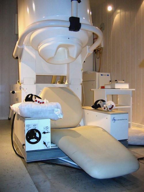
What does the equipment look like?
An MEG study is performed in a special room that is shielded from outside magnetic and electric noise. A helmet-shaped container is placed on the patient's head. Tiny magnetic sensors line the inside of this specially designed helmet, which looks similar to a large hair dryer. The computer workstation that helps detect and record the signals from the MEG helmet is located in a separate room.
How does the procedure work?
Brain cells interact by generating tiny electrical voltages that create electrical currents throughout the brain. This electrical flow produces magnetic fields that can be detected and recorded using sensitive magnetic sensors. Several hundred of these specialized sensors are built into the MEG helmet, which is placed on the patient's head. This sophisticated instrument and computer software work together to detect and record the activity of neurons as the patient lies still or completes a series of tasks, such as listening to a series of words or looking at pictures. An analysis of the recording, which collects both normal and abnormal brain signals every millisecond of the study, helps determine where specific activities in the brain originate.
How is the procedure performed?
MEG examinations are usually performed on an outpatient basis.
Typically, three to four positioning coils may be attached to your head with temporary tape to help precisely determine the location of your head relative to the MEG detectors.
An electroencephalogram (EEG), another type of test used to detect abnormalities related to the electrical activity of the brain, may be performed at the same time as the MEG exam. If so, small electrical conductors called electrodes will also be attached to your head with temporary tape, or on a cap that resembles a swimming-cap.
The exact positions of the coils and electrodes are measured using a special wand-like device called a digitizer.
The patient may be positioned on a moveable examination table or seated in a comfortable chair within a room that shields out any electric and magnetic noise that could interfere with the exam. You will be positioned within the stationary helmet that contains the MEG detectors placed on your head.
Depending on the type of study you are having, you may lie quietly or even go to sleep. If you are having an MEG exam to identify the sensory areas of your brain, you will be given ear phones and presented with sounds or images on a screen and asked to respond. To identify areas of the brain involved in movement, you may be asked to repeatedly push a button. For identifying language areas of the brain, you may be asked to read.
Throughout these tasks, you will be asked to hold relatively still and keep your head and eye movements to a minimum.
After your exam is finished, the recording will be analyzed.
MEG exams generally include multiple recording sequences, some of which may last several minutes.
The entire examination usually takes between one to two hours, depending on the extent of functional mapping.
What will I experience during and after the procedure?
Most MEG exams are painless.
Patients do not typically feel claustrophobic when wearing the MEG helmet because it fits loosely on the head and does not cover your face or body.
You will be alone in the exam room during much of the MEG procedure. At all times during your exam, the technologist will be able to see, hear and speak with you using a two-way intercom. Many MEG centers allow a friend or parent to stay in the room as long as they are also screened for safety in the magnetic environment.
If you or your child have been sedated or anesthetized for an MEG exam, recovery time ranges from approximately 30 minutes to two hours after the exam is completed.
If you have not been sedated, no recovery period is necessary. You may resume your usual activities and normal diet immediately after the exam.
Who interprets the results and how do I get them?
A radiologist, a physician specially trained to supervise and interpret radiology examinations, will analyze the images and send a signed report to your primary care or referring physician, who will share the results with you.
What are the benefits vs. risks?
Benefits
- MEG is a noninvasive imaging technique that does not involve exposure to ionizing radiation.
- MEG is a highly precise, real-time study of brain activity.
- MEG improves the surgical outcomes of epilepsy patients.
Risks
- The MEG examination poses no known risk to the average patient when appropriate safety guidelines are followed.
What are the limitations of MEG?
Patients need to remain relatively still during an MEG exam. Patients with a vagus nerve stimulator (VNS), pacemaker or similar device may not be able to undergo an MEG study.
This page was reviewed on April 15, 2022