Diagnostic Considerations
In most patients with erythema migrans, a carefully elicited history (including definitions of epidemiologic context) and a physical examination are all that is required to establish the diagnosis of Lyme disease. However, although many patients with Lyme disease present with erythema migrans, others first present with extracutaneous symptoms. In those cases, erythema migrans may never have occurred, may not have been recognized by the patient, or may not have been correctly diagnosed by the physician.
There can be a tendency to overdiagnose Lyme disease, especially in patients with a lifestyle that puts them in a high-risk category. Performing tests when the prior probability of disease is low increases the likelihood of false-positive results. The best way to avoid problems with diagnosis is to follow the Centers for Disease Control and Prevention (CDC) guidelines regarding diagnosis (see Workup), to use a reputable laboratory with experience in testing for Lyme disease, and to obtain the assistance of an infectious disease expert when any questions arise.
Because interpretation of testing is related to stage of disease and requires a two-stage test, laboratory results are often misinterpreted. Clinicians unfamiliar with Lyme disease or Lyme testing may falsely exclude the diagnosis by testing too early in the disease course, or falsely diagnose disease by following up negative enzyme immunoassay (EIA) results with Western blot testing (the latter is indicated only in patients with a positive or indeterminate EIA result).
In addition, separating false-positive antibody tests from asymptomatic infection is impossible. Approximately 5-10% of patients in endemic areas have positive antibody results without a history of symptoms.
Unfortunately, antibodies induced by the infection are not protective against further exposures to Borrelia burgdorferi; therefore, reinfection easily could be confused with a recurrence. Because antibodies may persist for years following an infection, repeat infection is a difficult diagnosis without specific signs of Lyme disease (eg, erythema migrans). Increasing titers after adequate treatment certainly raises suspicion of an active infection, but this is not a reason to repeat posttreatment titers, as they may remain positive for many years.
Other problems to be considered include the following:
-
Ankylosing spondylitis and rheumatoid arthritis
-
Atrioventricular (AV) nodal block
-
Cellulitis (see image below)Contact dermatitis
-
Granuloma annulare
-
Confusional states and acute memory disorders
-
Gout and pseudogout
-
Prion-related diseases
-
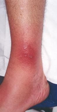 Lyme disease. The rash on the ankle seen in this photo is consistent with both cellulitis (deep red hue, acral location, mild tenderness) and erythema migrans (presentation in July, in an area highly endemic for Lyme disease). In this situation, treatment with a drug that covers both diseases (eg, cefuroxime or amoxicillin-clavulanate) is an effective strategy.

Co-infection
The pathogens responsible for babesiosis and ehrlichiosis share the same tick vector as B burgdorferi, making co-infection possible. Severe and even fatal acute infection caused by these pathogens is more common in asplenic individuals (babesiosis) or older adults (ehrlichiosis). Unlike B burgdorferi, however, these pathogens do not cause chronic infection. To add to the confusion, ehrlichial infection may cause a false-positive result for Lyme disease on immunoglobulin M (IgM) Western blot analysis.
In a prospective study from New York State, 52 adult patients with erythema migrans who had not received treatment for Lyme disease underwent testing for Anaplasma phagocytophilum, Babesia microti, Borrelia miyamotoi, and the deer tick virus subtype of Powassan virus. Nearly 90% of the patients showed no evidence of co-infection. Polymerase chain reaction (PCR) tests for B microti DNA were positive in 3 patients, one of whom also had a positive blood smear; these patients also had clinical signs suspicious for babesiosis. And additional 3 patients had elevated convalescent-phase IgG titers for B microti. [47]
The possibility of co-infection with another tick-borne pathogen should be considered if the patient’s condition does not respond to treatment as expected with ordinary early Lyme disease. Evidence suggests that co-infected patients have more symptoms of longer duration compared with other patients. In addition, these patients may be sicker than others on first observation.
Results of some laboratory studies may suggest some of the other co-infecting tick-borne pathogens such as ehrlichial or babesial species. Most patients with ehrlichiosis have elevated levels of hepatic transaminases, leukopenia, and/or thrombocytopenia. In addition, some patients have morulae (intracytoplasmic inclusions) in white blood cells, as demonstrated on peripheral blood smears.
Patients with babesiosis often are anemic (hemolytic type) and may have thrombocytopenia. Blood smears reveal the malarialike intraerythrocytic parasite in this disease as well, as shown below.
 Blood smear showing likely babesiosis. Babesiosis can be difficult to distinguish from malaria on a blood smear.
Differential Diagnoses
-
Lyme disease. The bacterium Borrelia burgdorferi (darkfield microscopy technique, 400X; courtesy of the US Centers for Disease Control and Prevention).
-
Lyme disease. Magnified ticks at various stages of development.
-
Ticks are the most common vectors for vector-borne diseases in the United States. In North America, tick bites can cause Lyme disease, human granulocytic and monocytic ehrlichiosis, babesiosis, relapsing fever, Rocky Mountain spotted fever, Colorado tick fever, tularemia, Q fever, and tick paralysis. Europe has a similar list of illnesses caused by ticks, but additional concerns include boutonneuse fever and tick-borne encephalitis. Lyme disease is one of the most prominent tick-borne diseases, and its main vector is the tick genus Ixodes, primarily Ixodes scapularis. Image courtesy of the US Centers of Disease Control and Prevention.
-
Lyme disease. Approximate US distribution of Ixodes scapularis. Image courtesy of the US Centers for Disease Control and Prevention.
-
Lyme disease. In general, Ixodes scapularis must be attached for at least 24 hours to transmit the spirochete to the host mammal. Prophylactic antibiotics are more likely to be helpful if feeding is longer. This photo shows 2 I scapularis nymphs. The one on the right is unfed; the other has been feeding for 48 hours. Note its larger size and the fact that the midgut diverticula (delicate brown linear areas on the body) are blurred. Photo by Darlyne Murawski; reproduced with permission.
-
Lyme disease. Normal and engorged Ixodes ticks.
-
Amblyomma americanum is the tick vector for monocytic ehrlichiosis and tularemia. An adult and a nymphal form are shown (common match shown for size comparison). Image by Darlyne Murawski; reproduced with permission.
-
Approximate US distribution of Amblyomma americanum. Image courtesy of the US Centers for Disease Control and Prevention.
-
The soft-bodied tick of the genus Ornithodoros transmits various Borrelia species that cause relapsing fever. Photo courtesy of Julie Rawlings, MPH, Texas Department of Health. Relapsing fever is characterized by recurrent acute episodes of fever (usually >39°C). It is a vector-borne illness spread by lice and ticks. The spirochete species Borrelia is responsible.
-
The Ixodes scapularis tick is considerably smaller than the Dermacentor tick. The former is the vector for Lyme disease, granulocytic ehrlichiosis, and babesiosis. The latter is the vector for Rocky Mountain spotted fever. This photo displays an adult I scapularis tick (on the right) next to an adult Dermacentor variabilis; both are next to a common match displayed for scale. Photo by Darlyne Murawski; reproduced with permission.
-
Approximate US distribution of Dermacentor andersoni. Image courtesy of the US Centers for Disease Control and Prevention.
-
Rhipicephalus ticks are vectors for babesiosis and rickettsial infections, among others. Image courtesy of Dirk M. Elston, MD. In typical practice, testing ticks for tick-borne infectious organisms is not generally recommended. However, healthcare practitioners should become familiar with the clinical manifestations of tick-borne diseases (eg, Lyme disease, especially those practicing in endemic areas) and maintain a high index of suspicion during warmer months. Ticks can be placed in a sealed container with alcohol if they need to be transported and identified.
-
To remove a tick, use fine-tipped forceps and wear gloves. Grasp the tick as close to the skin surface as possible, including the mouth parts, and pull upward with steady, even traction. Do not twist or jerk the tick because this may cause the mouth parts to break off and remain in the skin; however, note that the mouth parts themselves are not infectious. When removing, wear gloves to avoid possible infection.
-
Lyme disease. This patient's erythema migrans rash demonstrates several key features of the rash, including size, location, and presence of a central punctum, which can be seen right at the lateral margin of the inferior gluteal fold. Note that the color is uniform; this pattern probably is more common than the classic pattern of central clearing. On history, this patient was found to live in an endemic area for ticks and to pull ticks off her dog daily.
-
Erythema migrans, the characteristic rash of early Lyme disease.
-
Lyme disease. The thorax and torso are typical locations for erythema migrans. The lesion is slightly darker in the center, a common variation. In addition, this patient worked outdoors in a highly endemic area. Physical examination also revealed a right axillary lymph node.
-
Lyme disease. Photo of the left side of the neck of a patient who had pulled a tick from this region 7 days previously. Note the raised vesicular center, which is a variant of erythema migrans. The patient had a Jarisch-Herxheimer reaction approximately 18 hours after the first dose of doxycycline.
-
Lyme disease. Classic target lesion with concentric rings of erythema, which often show central clearing. Although this morphology was emphasized in earlier North American literature, it only represents approximately 40% of erythema migrans lesions in the United States. This pattern is more common in Europe. Courtesy of Lyme Disease Foundation, Hartford, Conn.
-
Typical appearance of erythema migrans, the bull's-eye rash of Lyme disease.
-
Lyme disease. Bulls-eye rash.
-
Lyme disease. Photo of erythema migrans on the right thigh of a toddler. The size and location are typical of erythema migrans, as is the history of the patient vacationing on Fire Island, NY, in the month of August. No tick bite had been noted at this location. Approximately 25% of patients with Lyme disease are children, which is the same percentage of patients who do not recall a tick bite. Courtesy of Dr John Hanrahan.
-
Lyme disease. Multiple lesions of erythema migrans occur in approximately 20% of patients. A carpenter from Nantucket who worked predominantly outside had been treated with clotrimazole/betamethasone for 1 week for a presumed tineal infection, but the initial lesion grew, and new ones developed. He then presented to the emergency department with the rashes seen in this photo. The patient had no fever and only mild systemic symptoms. He was treated with a 3-week course of oral antibiotics.
-
Lyme disease. The rash on the ankle seen in this photo is consistent with both cellulitis (deep red hue, acral location, mild tenderness) and erythema migrans (presentation in July, in an area highly endemic for Lyme disease). In this situation, treatment with a drug that covers both diseases (eg, cefuroxime or amoxicillin-clavulanate) is an effective strategy.
-
Lyme disease. Borrelial lymphocytoma of the earlobe, which shows a bluish red discoloration. The location is typical in children, as opposed to the nipple in adults. This manifestation of Lyme disease is uncommon and occurs only in Europe. Courtesy of Lyme Disease Foundation, Hartford, Conn.
-
A rarely reported noninfectious complication for tick bites is alopecia. It can begin within a week of tick removal and typically occurs in a 3- to 4-cm circle around a tick bite on the scalp. A moth-eaten alopecia of the scalp caused by bites of Dermacentor variabilis (the American dog tick) has also been described. No particular species appears more likely to cause alopecia. Hair regrowth typically occurs within 1-3 months, although permanent alopecia has been observed.
-
Acrodermatitis chronica atrophicans is found almost exclusively in European patients and comprises an early inflammatory phase and a later atrophic phase. As the term suggests, the lesion occurs acrally and ultimately results in skin described as being like cigarette paper. Courtesy of Lyme Disease Foundation, Hartford, Conn.
-
Blood smear showing likely babesiosis. Babesiosis can be difficult to distinguish from malaria on a blood smear.
-
Life cycle of the Ixodes dammini tick. Courtesy of Elsevier.
-
Lyme disease in the United States is concentrated heavily in the northeast and upper Midwest; it does not occur nationwide. Dots on the map indicate the infected person's county of residence, not the place where they were infected. Courtesy of the US Centers for Disease Control and Prevention (CDC).
Tables
- Table 1. Clinical presentation and therapy for the stages of Lyme disease
- Table 2. Adult and pediatric treatment options, dosages, and routes of administration
- Table 3. Comparison of Infectious Diseases Society of America (IDSA) and International Lyme and Associated Diseases Society (ILADS) recommendations for Lyme disease treatment
Clinical Manifestations |
Adult Dose |
Pediatric Dose |
Erythema migrans |
Doxycycline 100 mg PO BID for 10-14 days, OR
Amoxicillin 500 mg PO TID for 14 days, OR
Cefuroxime axetil 500 mg PO BID for 14 days
Patients unable to take doxycycline or beta-lactam antibiotics: Azithromycin 500 mg PO qDay for 7 days |
Doxycycline 4.4 mg/kg/day PO, divided into 2 doses; not to exceed 100 mg/dose for 10-14 days, OR
Amoxicillin 50 mg/kg/day PO, divided into 3 doses; not to exceed 500 mg/dose for 14 days, OR
Cefuroxime axetil 30 mg/kg/day PO, divided into 2 doses; not to exceed 500 mg/dose for 14 day |
Facial palsy [3] |
Doxycycline 100 mg PO BID for 14-21 days |
Doxycycline 4.4 mg/kg/day PO, divided into 2 doses; not to exceed 100 mg/dose for 14-21 days |
Lyme meningitis or radiculoneuritis [3] |
Doxycycline 200 mg/day PO, divided into 1-2 doses for 14-21 days, OR
Ceftriaxone 2 g IV qDay for 14-21 days; may substitute for oral therapy once the patient is stabilized or discharged to complete the course |
Doxycycline 4.4 mg/kg/day PO, divided into 1-2 doses; not to exceed 100 mg/dose for 14-21 days, OR
Ceftriaxone 50-75 mg/kg IV qDay; not to exceed 2 g/day for 14-21 days; may substitute for oral therapy once the patient is stabilized or discharged to complete the course |
Lyme disease–associated meningitis, cranial neuropathy, radiculoneuropathy, or with other peripheral nervous system (PNS) manifestations |
Without parenchymal involvement of brain or spinal cord: IV ceftriaxone, cefotaxime, penicillin G, or oral doxycycline With parenchymal involvement of brain or spinal cord: IV antibiotics are preferred |
|
Mild lyme carditis (1st degree AV block with PR interval < 300 milliseconds) [3] |
Doxycycline 100 mg PO BID for 14-21 days, OR
Amoxicillin 500 mg PO TID for 14-21 days, OR Cefuroxime axetil 500 mg PO BID for 14-21 days |
Doxycycline 4.4 mg/kg/day PO, divided into 2 doses; not to exceed 100 mg/dose for 14-21 days, OR
Amoxicillin 50 mg/kg/day PO, divided into 3 doses; not to exceed 500 mg/dose for 14-21 days, OR
Cefuroxime axetil 30 mg/kg/day PO, divided into 2 doses; not to exceed 500 mg/dose for 14-21 days |
Severe lyme carditis (symptomatic, 1st degree AV block with PR interval ≥300 milliseconds, 2nd or 3rd degree AV block) [3] |
Ceftriaxone 2 g IV qDay for 14-21 days; once symptoms and high-grade AV block resolve, consider transitioning to oral antibiotics to complete treatment course |
Ceftriaxone 50-75 mg IV qDay; not to exceed 2 g/day for 14-21 days; once symptoms and high-grade AV block resolve, consider transitioning to oral antibiotics to complete treatment course |
Borrelial lymphocytoma |
Oral antibiotic therapy for 14 days |
|
Arthritis |
Doxycycline 100 mg PO BID for 28 days, OR
Amoxicillin 500 mg PO TID for 28 days, OR
Cefuroxime axetil 500 mg PO BID for 28 days |
For ≥8 years:
Doxycycline 4.4 mg/kg/day PO, divided into 2 doses; not to exceed 100 mg/dose for 28 days, OR
Amoxicillin 50 mg/kg/day PO, divided into 3 doses; not to exceed 500 mg/dose for 28 days, OR
Cefuroxime axetil 30 mg/kg/day PO, divided into 2 doses; not to exceed 500 mg/dose for 28 days
For < 8 years:
Amoxicillin 50 mg/kg/day PO, divided into 3 doses; not to exceed 500 mg/dose for 28 days, OR
Cefuroxime axetil 30 mg/kg/day PO, divided into 2 doses; not to exceed 500 mg/dose for 28 days |
Arthritis without any response to initial treatment |
Ceftriaxone 2 g IV qDay for 14-28 days |
Ceftriaxone 50-75 mg IV qDay; not to exceed 2 g/day for 14-28 days |
Acrodermatitis chronica atrophicans |
Oral antibiotic therapy 21-28 days |
|
|
Treatment |
Adult Dose |
Pediatric Dose |
Oral Therapy |
Doxycycline (patients > 8 y) |
100 mg twice a day |
Doxycycline 4.4 mg/kg/day PO (up to 100 mg BID) |
Amoxicillin |
500 mg three times a day |
50 mg/kg/day (up to 500 mg) in 3 divided doses |
|
Cefuroxime axetil |
500 mg twice a day |
30 mg/kg/day (up to 500 mg) in 2 divided doses |
|
Phenoxymethylpenicillin |
500 mg four times a day, or 1 gm three times a day |
50-100 mg/kg/day in three divided doses; maximum 1 g/dose |
|
Azithromycin (for patients unable to take doxycycline or beta-lactams) |
500 mg once a day
|
50-100 mg/kg/day in three divided doses; maximum 1 g/dose |
|
Intravenous therapy |
Ceftriaxone |
2 g once a day |
10 mg/kg/day (maximum, 500 mg/day) |
Cefotaxime |
2 g every 8 h |
150-200 mg/kg (up to 2 g) every 8 h |
|
Penicillin G |
18-24 million U/d divided every 4 h |
200,000-400,000 mg/kg (up to 2 g) every 8 h |
Treatment Focus |
IDSA |
ILADS |
Treatment of a tick bite without symptoms of Lyme disease |
Doxycycline, 200 mg as a single dose |
Doxycycline, 100 mg bid for 20 days |
Erythema migrans |
Doxycycline, amoxicillin, or cefuroxime for 14-21 days |
Doxycycline, amoxicillin, or cefuroxime for 28-42 days or azithromycin for at least 21 days |
“Persisting symptoms of Lyme disease” |
No antibiotic therapy |
Multiple agents (individually or in combination) are mentioned without specific doses or duration recommended |