Lateral plantar nerve
Last updated| Lateral plantar nerve | |
|---|---|
 The plantar nerves. | |
 | |
| Details | |
| From | Tibial nerve |
| Innervates | sole, abductor digiti minimi muscle (foot), flexor digiti minimi brevis muscle (foot), quadratus plantae, 3 lateral lumbricals of the foot, adductor hallucis muscle, plantar interossei muscles, dorsal interossei muscles |
| Identifiers | |
| Latin | Nervus plantaris lateralis |
| TA | A14.2.07.069 |
| FMA | 44724 |
| Anatomical terms of neuroanatomy | |
The lateral plantar nerve (external plantar nerve) is a branch of the tibial nerve, in turn a branch of the sciatic nerve and supplies the skin of the fifth toe and lateral half of the fourth, as well as most of the deep muscles, its distribution being similar to that of the ulnar nerve in the hand.

The tibial nerve is a branch of the sciatic nerve. The tibial nerve passes through the popliteal fossa to pass below the arch of soleus.

The sciatic nerve is a large nerve in humans and other animals. It begins in the lower back and runs through the buttock and down the lower limb. It is the longest and widest single nerve in the human body, going from the top of the leg to the foot on the posterior aspect. The sciatic nerve provides the connection to the nervous system for nearly the whole of the skin of the leg, the muscles of the back of the thigh, and those of the leg and foot. It is derived from spinal nerves L4 to S3. It contains fibers from both the anterior and posterior divisions of the lumbosacral plexus.
Toes are the digits of the foot of a tetrapod. Animal species such as cats that walk on their toes are described as being digitigrade. Humans, and other animals that walk on the soles of their feet, are described as being plantigrade; unguligrade animals are those that walk on hooves at the tips of their toes.
Contents
It passes obliquely forward with the lateral plantar artery to the lateral side of the foot, lying between the flexor digitorum brevis and quadratus plantae and, in the interval between the flexor muscle and the abductor digiti minimi, divides into a superficial and a deep branch. Before its division, it supplies the quadratus plantae and abductor digiti minimi. It divides into deep and superficial branches.

The lateral plantar artery, much larger than the medial, passes obliquely lateralward and forward to the base of the fifth metatarsal bone.
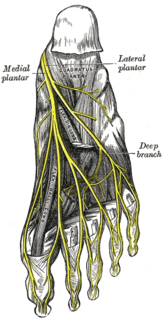
The deep branch of lateral plantar nerve accompanies the lateral plantar artery on the deep surface of the tendons of the Flexor muscles and the Adductor hallucis, and supplies all the Interossei, the second, third, and fourth Lumbricales.
The superficial branch of the lateral plantar nerve splits into a proper and a common plantar digital nerve:
Additional images

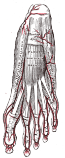
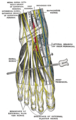 Nerves of the dorsum of the foot.


A coronal plane is any vertical plane that divides the body into ventral and dorsal sections.
Related Research Articles

The human leg, in the general meaning, is the entire lower limb of the human body, including the foot, thigh and even the hip or gluteal region. However, the definition in human anatomy refers only to the section of the lower limb extending from the knee to the ankle, also known as the crus. Legs are used for standing, and all forms of locomotion including recreational such as dancing, and constitute a significant portion of a person's mass. Female legs generally have greater hip anteversion and tibiofemoral angles, but shorter femur and tibial lengths than those in males.

The median nerve is a nerve in humans and other animals in the upper limb. It is one of the five main nerves originating from the brachial plexus.

In human anatomy, the ulnar nerve is a nerve that runs near the ulna bone. The ulnar collateral ligament of elbow joint is in relation with the ulnar nerve. The nerve is the largest unprotected nerve in the human body, so injury is common. This nerve is directly connected to the little finger, and the adjacent half of the ring finger, innervating the palmar aspect of these fingers, including both front and back of the tips, perhaps as far back as the fingernail beds.

The opponens digiti minimi is a muscle in the hand. It is of a triangular form, and placed immediately beneath the palmaris brevis, abductor digiti minimi and flexor digiti minimi brevis. It is one of the three hypothenar muscles that control the little finger.
The flexor digitorum brevis lies in the middle of the sole of the foot, immediately above the central part of the plantar aponeurosis, with which it is firmly united.

The quadratus plantae is separated from the muscles of the first layer by the lateral plantar vessels and nerve. It acts to aid in flexing the 2nd to 5th toes and is one of the few muscles in the foot with no homolog in the hand.

The plantar nerves are a pair of nerves innervating the sole of the foot. They arise from the posterior branch of the tibial nerve.
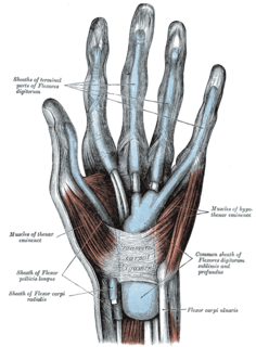
The hypothenar muscles are a group of three muscles of the palm that control the motion of the little finger.
The abductor digiti minimi is a muscle which lies along the lateral (outer) border of the foot, and is in relation by its medial margin with the lateral plantar artery, vein and nerves.
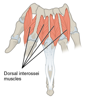
In human anatomy, the dorsal interossei (DI) are four muscles in the back of the hand that act to abduct (spread) the index, middle, and ring fingers away from hand's midline and assist in flexion at the metacarpophalangeal joints and extension at the interphalangeal joints of the index, middle and ring fingers.

In human anatomy, the abductor digiti minimi is a skeletal muscle situated on the ulnar border of the palm of the hand. It forms the ulnar border of the palm and its spindle-like shape defines the hypothenar eminence of the palm together with the skin, connective tissue, and fat surrounding it. Its main function is to pull the little finger away from the other fingers.

The flexor digiti minimi brevis is a hypothenar muscle in the hand that flexes the little finger at the metacarpophalangeal joint. It lies lateral to the abductor digiti minimi when the hand is in anatomical position.
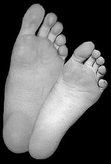
The sole is the bottom of the foot.
The medial plantar nerve is the larger of the two terminal divisions of the tibial nerve, which accompanies the medial plantar artery.

The deep branch of the ulnar nerve is a terminal, primarily motor branch of the ulnar nerve. It is accompanied by the deep palmar branch of ulnar artery.

The cervical spinal nerve 8 (C8) is a spinal nerve of the cervical segment.

The sacral spinal nerve 1 (S1) is a spinal nerve of the sacral segment.
References
This article incorporates text in the public domain from page 963 of the 20th edition of Gray's Anatomy (1918)
The public domain consists of all the creative works to which no exclusive intellectual property rights apply. Those rights may have expired, been forfeited, expressly waived, or may be inapplicable.

Gray's Anatomy is an English language textbook of human anatomy originally written by Henry Gray and illustrated by Henry Vandyke Carter. Earlier editions were called Anatomy: Descriptive and Surgical, Anatomy of the Human Body and Gray's Anatomy: Descriptive and Applied, but the book's name is commonly shortened to, and later editions are titled, Gray's Anatomy. The book is widely regarded as an extremely influential work on the subject, and has continued to be revised and republished from its initial publication in 1858 to the present day. The latest edition of the book, the 41st, was published in September 2015.
External links
Text is available under the CC BY-SA 4.0 license; additional terms may apply.
Images, videos and audio are available under their respective licenses.