

- 📖 Geeky Medics OSCE Book
- ⚡ Geeky Medics Bundles
- ✨ 1300+ OSCE Stations
- ✅ OSCE Checklist PDF Booklet
- 🧠 UKMLA AKT Question Bank
- 💊 PSA Question Bank
- 💉 Clinical Skills App
- 🗂️ Flashcard Collections | OSCE, Medicine, Surgery, Anatomy
- 💬 SCA Cases for MRCGP
To be the first to know about our latest videos subscribe to our YouTube channel 🙌
Introduction
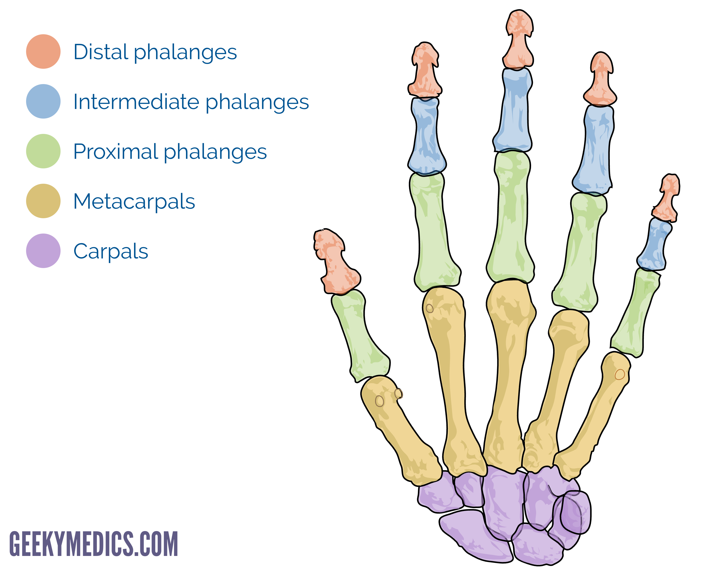
Anatomically, the hand is defined as the region of the upper limb distal to the wrist.
The base of the hand contains 8 bones, each known as a carpal bone. The palms of the hands each contain 5 metacarpal bones. The digits contain the phalanges.
The skeleton of the hand contains 27 bones which can be divided into three groups:
- The carpus (the wrist): comprised of 8 carpal bones
- The metacarpus: comprised of 5 metacarpal bones
- The phalanges: comprised of 14 phalangeal bones
Approximately 1.8 million people attend Emergency Departments each year due to hand injuries.1 The functional impact of an injury to the hand is often out of proportion with the extent of the injury itself. As a result, it is important to have a good understanding of the anatomy of the hand.
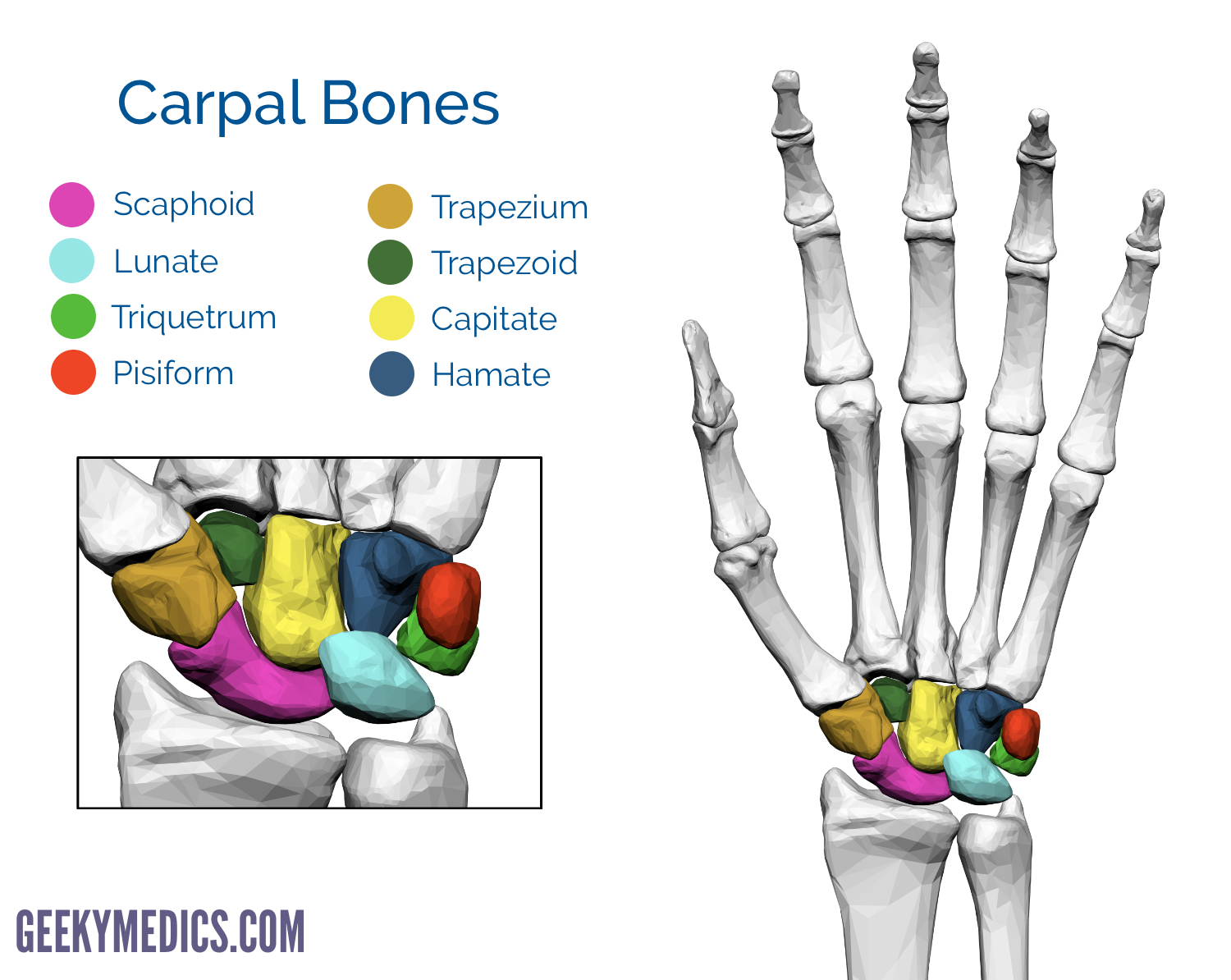
The carpal bones
The 8 carpal bones are arranged into two rows of four: a proximal and a distal row.
The proximal row of carpal bones, from the lateral (thumb side) to the medial side is made up of the following bones:
- Scaphoid
- Lunate
- Triquetrum
- Pisiform
The distal row of carpal bones, laterally to medially is made up of the following bones:
- Trapezium
- Trapezoid
- Capitate
- Hamate
Mnemonic
A useful mnemonic to help remember the carpal bones is shown below:
- Some – Scaphoid
- Lovers – Lunate
- Try – Triquetrum
- Positions – Pisiform
- That – Trapezium
- They – Trapezoid
- Can’t – Capitate
- Handle – Hamate
Scaphoid
Scaphoid (“boat-shaped”) is a boat-shaped bone which articulates proximally with the radius to form the radial border of the carpal tunnel.
The scaphoid is the largest bone in the proximal row of carpal bones and is also the most commonly fractured. It often occurs due to a fall on an outstretched hand. As a result of the poor blood supply to the scaphoid, fractures can be slow to heal and avascular necrosis of the proximal fragment of the scaphoid can occur.
Lunate
Lunate (“moon-shaped”) is a crescent-shaped bone articulating proximally with the radius. The lunate is found centrally in the carpal bones between the scaphoid and triquetrum. The lunate bone is the most frequently dislocated carpal bone.
Triquetrum
Triquetrum (“three-cornered”) is a pyramidal bone. The triquetrum lies between the lunate and pisiform bones on the medial side of the proximal row of carpal bones. Fractures of the triquetrum often occur upon forceful dorsiflexion of the hand such that an avulsion fracture occurs on the dorsal aspect of the bone.
Pisiform
Pisiform (“pea-shaped”) is a small round sesamoid bone found in the tendon of the flexor carpi ulnaris. The pisiform articulates with the anterior surface of the triquetrum bone, therefore extending anteriorly to form the bump that can be felt at the medial base of the hand. The pisiform bone forms the ulnar border of the carpal tunnel. In contrast to the other carpal bones, the pisiform is not involved in movements of the wrist.
Trapezium
Trapezium (“table”) is a four-sided bone. It articulates with four bones; the 1st and 2nd metacarpals, the scaphoid and the trapezoid. The trapezium has a distinct tubercle on the palmar surface which projects anteriorly.
Trapezoid
The trapezoid (“resembles a table”) is a wedge-shaped bone. It gets its name due to its similarity to the trapezium. The trapezoid is the smallest bone in the distal row of carpal bones. The trapezoid articulates with four bones; the 2nd metacarpal, the trapezium, the capitate and the scaphoid bone. The trapezoid is the least commonly fractured carpal bone.
Capitate
Capitate (“head-shaped”) is a head-shaped bone. The capitate is the largest carpal bone and articulates with five bones; the 3rd metacarpal, the trapezoid, scaphoid, lunate and hamate.
Hamate
Hamate (“hooked bone”) is a wedge-shaped bone. It articulates with five carpal bones; the 4th and 5th metacarpals, the capitate, and the triquetrum. The hamate is easily distinguishable due to its shape and a hook-like process that extends towards the palmar surface.
The hamate bone is the most frequently fractured bone when a golfer hits the surface hard with a golf club on the downswing. Clinically, this may present with pain and symptoms of ulnar nerve damage; numbness and weakness of flexion in the 4th and 5th fingers. In a similar fashion to the scaphoid, the hook of the hamate is vulnerable to avascular necrosis due to its poor blood supply.
The carpal tunnel
The carpal bones form a U-shaped arrangement which is directed anteriorly. The flexor retinaculum spans this U-shaped area to maintain the alignment of the carpal bones.
The medial side of the base of the arch is formed by the pisiform and hook of the hamate. The lateral side of the base of the arch is formed by the scaphoid and trapezium.
The flexor retinaculum attaches laterally to the trapezium and scaphoid bones and medially to the hamate and pisiform bones. Both the carpal bones and the flexor retinaculum form the carpal tunnel– the bones forming the wall and floor and the tendon, the roof.
Thickening of the flexor retinaculum can result in carpal tunnel syndrome which results in symptoms secondary to compression of the median nerve.
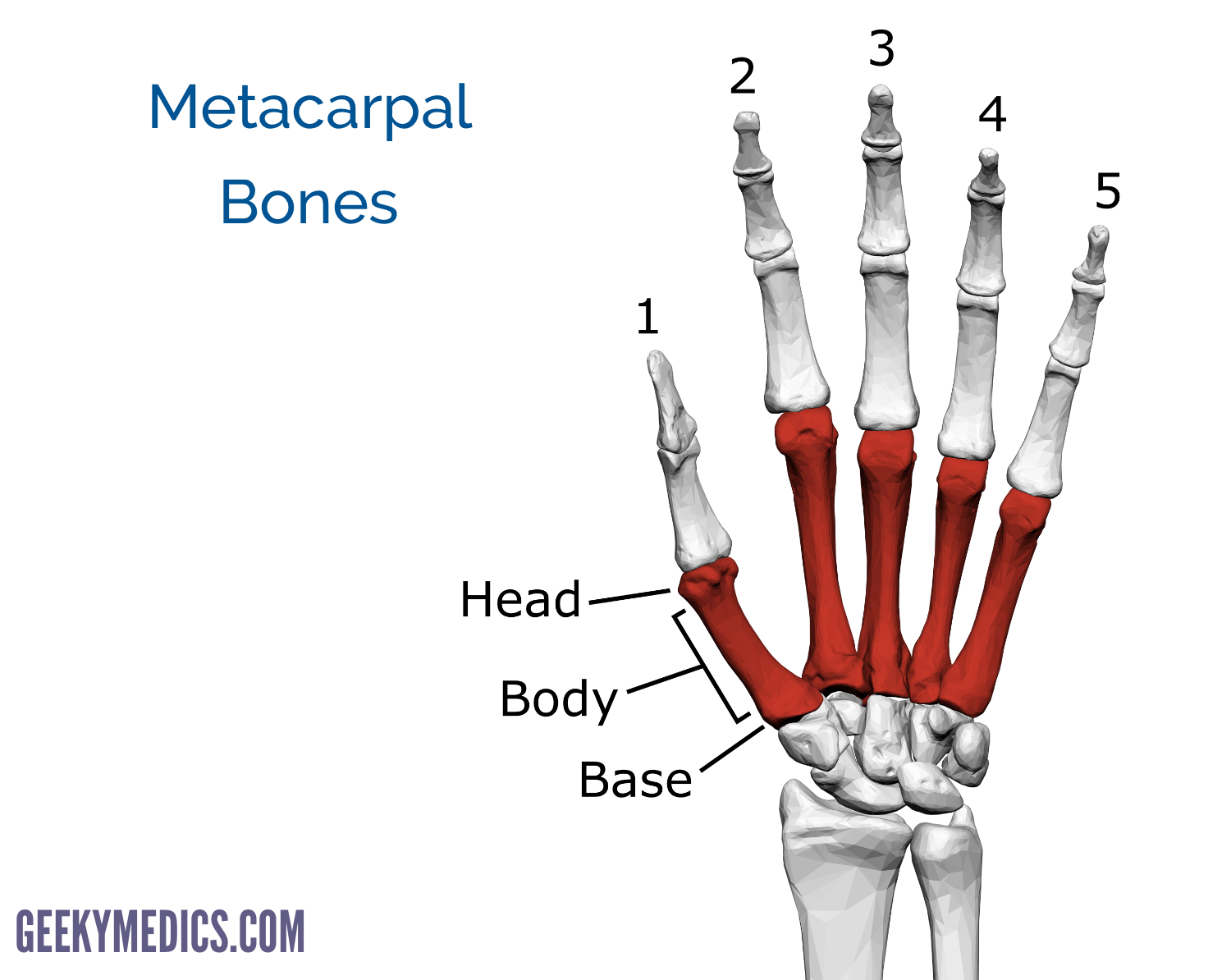
Metacarpal bones
Each palm contains 5 metacarpals bones which articulate proximally with one of the distal carpal bones forming a carpometacarpal joint. The distal end of the metacarpals articulate with the phalanges forming a metacarpophalangeal joint.
Each metacarpal is related to a digit and numbered 1-5, beginning with the thumb. Each metacarpal has a head, body and a base. The knuckles are formed by the heads of the metacarpals.
The first metacarpal bone is separated from all the other metacarpal bones which form the base of the hand. This allows free movement of the first metacarpal, independent of all the other metacarpals bones.
As metacarpals 2-5 are articulated closely together, fractures of the metacarpals tend to be stable, however, instability can occur should multiple metacarpal fractures occur (e.g. in a severe crush trauma). The neck of the 4th and 5th metacarpals is a common location for a boxer’s fracture, this classically occurs when an individual hits an object with a closed fist.
Phalanges
There are 14 phalanges which make up the bones of the fingers and thumbs. Similar to the hallux, the Pollux (thumb) only has two phalanges (proximal and distal) whereas the remaining digits have three (proximal, middle and distal).
Like the metacarpals, each phalanx has a base, body and distally, a head. The base of each phalanx articulates with the head of the metacarpal.
The most common type of injury to the phalanges are crushing injuries, for example, closing a door on the digit.
Reviewer
Mr Saleem Mastan
Trauma & Orthopaedic Registrar
References
- Murphy GRF, Gardiner MD, Glass GE, Kreis IA, Jain A, Hettiaratchy S. Meta-analysis of antibiotics for simple hand injuries requiring surgery. Br J Surg [Internet]. 2016 Apr;103(5):487–92.
- By BodyParts3D is made by DBCLS. Rendering the image is by was_a_bee, using Blender. – Polygon data is from BodyParts3D. License: [CC BY-SA 2.1]. Available from: [LINK]
