
Name
Greek: entos = inner, amoibos = changing shape, histos = tissue, and lysis = dissolution of tissues.
Geographic Distribution/Epidemiology
These amoebae are found in warm countries all over the world, but are also often imported by travelers and refugees into temperate climatic zones (Fig. 1). More than 500 million humans are constantly infected. Death tolls per year are calculated 75,000–100,000. About 10 % of the world population is constantly endangered.


Distribution map of amoebic dysenteria (WHO)
Morphology/Life Cycle
This amoeba may occur in three different stages inside the intestine of humans (Figs. 2, 3, 4, and 5).


Life cycle of Entamoeba histolytica. 1 Cysts with 4 nuclei (i.e., metacysts) are ingested orally with contaminated food or drinking water (a-c). 2–4 After excysting in the small intestine, both the cytoplasm and nuclei divide to form 8 small amoebulae (i.e., metacystic trophozoites). 5,6 Mature trophozoites (i.e., minuta forms) reproduce by constant binary fissions. 7 Uninucleate cyst (i.e., precyst) contains chromatoid bodies and (often) a large glycogen vacuole. 8 Cysts with 2 nuclei and chromatoid bodies. 9 Cysts with 4 nuclei (metacysts) are set free with the feces and become infectious when ingested by man. 10–11 Some of the minuta forms may grow to magna forms, which enter the intestinal wall and, via the bloodstream, other organs such as the liver, lung, and brain (11a-c), where they lead to abscesses (i.e., ameboma). Living amoebae are only found at the periphery of these ameboma. (ABabscess; CH chromatoid body; CW cyst wall; E erythrocyte; P single, pale pseudopodium; N nucleus with central nucleolus (karyosome); NV food vacuole; V glycogen vacuole of young cysts (for further species, see Amoebae/Table 1)


Unstained minuta stage of E. histolytica. Note the centrally located nucleolus in the nucleus


Light micrographs of a minuta stage amoeba (a), magna form containing red blood cells (b), and a cyst (c) containing the 4 nuclei (with each a central nucleolus); N = nucleus; W = cyst wall


Two magna forms seen by help of Nomarski contrast
-
(a)
Minuta stage (10–20 μm)
-
(b)
Magna stage (20–40 μm)
-
(c)
Cyst stage (10–15 μm)
Minuta and magna stages are characterized by a hyaline ectoplasm, a single hernia sack-like protruding pseudopodium at the anterior pole of the cell and as seen in colored specimens by their typical nucleus and by condensation of the chromatin at the inner side of the inner nuclear membrane (Figs. 2, 3, and 4). The centrally situated nucleolus is smaller than that of E. gingivalis. The minuta stage lives in the intestinal fluid and divides by binary fission. Feeding occurs by phagocytosis of particles from the intestinal fluid. When being transported in the region of the colon (where water retraction occurs), the minuta stages excrete a cyst wall and this stage now appears spherical (Figs. 2, 4c). The nucleus inside the young cyst divides and each of the progeny divides again once, so that cysts excreted with the feces each contain four nuclei all showing the typical central nucleolus (Figs. 2, 4c). Even completely unsymptomatic, persons may excrete 30–40 millions of these cysts per day in endemic regions. Since normal light microscopic investigations do not show differences between the newly described nonpathogenic species Entamoeba dispar and E. histolytica, it must be suggested that these reports of the excretion of millions of cysts are mainly based on infections with E. dispar and/or on apparently existing nonpathogenic strains of E. histolytica. It is estimated that E. dispar covers about 90 % of the excreted cysts. Recently it was shown that E. nuttalli cysts found in macaques are genetically different from E. histolytica, but appear identical.
Under certain conditions not well understood even today, the minuta stages may switch into magna stages (Fig. 5), which are able by excretion of substances (amoebapora) to attach at the cells of the intestinal wall, to destroy them, to find a way to enter blood vessels, and thus to reach finally the liver, where they form so-called liver abscesses (ameboma), which are filled by a brownish fluid consisting of bacteria, remnants of host cells, and rather few amoebae. These abscesses can easily be seen in computer-tomographic images, reaching diameters of 12–15 cm and may become life-threatening (Fig. 7).
Symptoms of Disease
The disease due to amoebic stages which lyse – according to their species name histolytica – liver tissues is called in literature entamoebiasis, amoebic dysenteria, or red dysenteria. There are distinguished two different phases:
-
(a)
Intestinal amoebiasis: Shortly after the infection, the patients complain light gastrointestinal symptoms such as feeling of abdominal pressure, wandering pain along the intestine, symptoms of vomiting, but never fever. The sudden onset of fever would be a sign of an infection with bacteria (bacterial dysenteria). At the beginning of an intestinal amoebiasis, the stool is formed or slightly pasty/mushy and contains whitish slime components. Ongoing with the infection, the number of defecations increases up to 5–10 per day (there are even more in cases of bacterial dysenterias!). In case of amoebic dysenteria, feces are only rarely fully watery, but contain in most cases bloody traces and larger amounts of slime, appearing like raspberry jelly (Fig. 6), while bacterial dysenterias appear yellowish-whitish purulently. During ongoing intestinal entamoebiasis, the general fitness of the patients becomes reduced. Ulcerations of the colon wall grow in number and start to confluence, while the so-called bottleneck and buttonhole ulcers are formed. Their borders are evaginated and their depth often reaches into the muscularis propria. These invaginations become quickly filled by huge amounts of bacteria. Depending on the size, localization, and number of these ulcers, the patients suffer from colic pain, decreasing fitness, and intermittent high fever. Since blood vessels are often destroyed inside the ulcers, the dysenteric stools contain increasing amounts of blood (Fig. 6). In case arteries become lysed, life-threatening bleedings may occur, which require surgical actions. All these symptoms resemble strongly the symptoms of a colitis ulcerosa; however, magna stages of Entamoeba histolytica do not occur there.
Entamoeba histolytica, Fig. 6 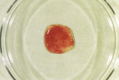
Portion of bloody stool
As complication of the intestinal entamoebiasis, a so-called intestinal ameboma structure may occur, which is a locally limited tumor, which is of the benign type but is also able to block (at least in parts) the discharge of the feces. Further complications such as perforation of the intestine and peritonitis may occur as a consequence. In several untreated cases, the intestinal symptoms may disappear; however, there is no guarantee that all infectious stages had disappeared.
-
(b)
Extraintestinal amoebiasis: This disease does not necessarily take a dramatic course. Very often slight abdominal pain and increased numbers of defecations remain for long the only symptoms. Also the examination of the blood does not show much pecularities. This slow progress of the symptoms, however, does not exclude the hidden formation of abscesses in the liver, but also in the lung or even (rarely!) in the brain. This phase of the early formation of an abscess may start only after months or even after years and is characterized by weakness, fever around 38–39 °C (usually without ague), and feeling of increasing local pain due to formation of cavities in tissues. With respect to the correct medical term, E. histolytica does not form true “abscesses,” since there is no limiting membrane or a wall around the hollow that occurs in the organs due to lysis of the tissues. Most Entamoeba abscesses are situated inside the right liver portion and initiate due to their quick and intense increase spontaneous pressure pain in the region of the upper right belly. In case the abscess is situated close to the diaphragm, it leads to sting-like pain during breathing. The patient lays rightward bended, breathes only slightly, and refuses touching. Occasionally an infiltration is formed by remnants of an inflammation along the right thorax wall. These symptoms are connected with an extremely high leukosis in the peripheral blood (reaching often values of more than 20,000/μl). The quickly increasing erythrocyte sedimentation rate (ESR) is a leading symptom for the existence of an acute liver abscess (ameboma) due to E. histolytica (Fig. 7). Investigations using ultrasonic waves or by help of computer tomography will confirm the presence of such abscesses. As soon as treatment is done, the symptoms of the disease are quickly reduced. Already on the following day of treatment, pain, fever, liver swelling, and inflammation symptoms decrease considerably. If this relief does not occur, the abscess has probably another origin and might be based on a tumor, on infections with bacteria, or on an infection with Echinococcus cysts. Depending on the size of the abscess, chirurgical intervention (e.g., sucking off the fluid of the abscess) may be needed but has to be done at all caution in order to avoid penetration of amoebae into the peritoneum. The surgeon has to decide whether intervention is needed or whether application of medicaments is recommended.
Entamoeba histolytica, Fig. 7 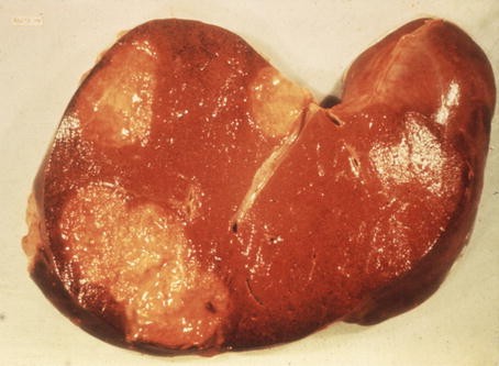
Section through a liver with several abscesses (ameboma)


Diagnosis
In the case of an intestinal entamoebiasis, cysts and trophozoites (minuta forms) can be microscopically diagnosed in material obtained from stools or by help of endoscopy. Cyst stages of E. histolytica can be demonstrated when using methods of fecal enrichment (M.I.F.C., S.A.F.C.; Fig. 4c). Modern PCR techniques may significantly differentiate between pathogenic and nonpathogenic species. However, motile magna forms would show their single pseudopodium only in fresh and body warm material. But the size (20–40 μm) and the engorged erythrocytes in those amoebae are characteristic and basis of a sure diagnosis. If an immediate investigation is not possible, the obtained material must be adequately preserved, e.g., in PVA, M.I.F.C., or S.A.F.C. solutions. In colored preparations (Heidenhain, trichrome, or Lawless), which were prepared from fresh or PVA-fixed material, the trophozoites of Entamoeba histolytica can be significantly differentiated from apathogenic intestinal amoebae by help of the structure of their nuclei. Sections of the intestinal wall also would show magna forms (Fig. 5). Stool from patients with perforated intestinal wall shows bloody contents (Fig. 6).
Extraintestinal amoebiasis is proven by the help of serodiagnostic techniques (IFT, ELISA) or by PCR, but to prove is not even easy at the beginning, and thus tests have to be repeated several times. However, in cases where the abscess has reached a certain size, computer tomography or sonograms are the techniques of choice (Fig. 7).
Prophylaxis
Avoid eating unwashed or non-peeled fruits or non-washed salads. Drinking water has to be cooked in endemic regions. Flies have to be kept off from kitchen or stored food.
Incubation Period
Two to 21 days; formation of abscesses in the liver and other organs may start about 1–3 months after an intestinal infection.
Prepatent Period
Two to seven days.
Patency
Eventually years.
Treatment
Medicaments of choice are nitroimidazoles, since they act as well against stages in the intestine as against amoebic stages disseminated in different organs. For example, metronidazole (3 × 500 -700 mg daily for 5–10 days for adults or 30 mg/kg body weight daily for 5–10 days) has very good curative effects. However, after metronidazole treatment, cyst excretion may persist, although there are no more symptoms of disease. These cases can be cured by application of diloxanide (3 × 500 mg daily oral for 10 days) or paromomycin (3 × 500 mg daily oral for 8–10 days). In cases of bleeding due to perforations chirurgical intervention is needed. Attention: Do not use metronidazole or paromomycin during pregnancy.
Further Reading
Anuar TS et al (2012) Molecular epidemiology of amoebiasis in Malaysia: highlighting the different risk factors of Entamoeba histolytica and E. dispar. Int J Parasitol 42:1165–1175
Anuar TS et al (2013) Evaluation of formalin-ether sedimentation and trichome staining techniques: its effectiveness in detecting Entamoeba histolytica/dispar/moshkovskii in stool samples. J Microbiol Methods 92:344–348
Choudhuri G, Rangan M (2012) Amebic infection in humans. Ind J Gastroenterol 32:153–162
Guzman-Silva MA et al (2013) Experimental amoebic liver abscess in hamsters caused by trophozoites of a Brazilian strain of Entamoeba dispar. Exp Parasitol 134:39–47
Haider SS et al (2013) Blastocystis spp., Cryptosporidium spp. and Entamoeba histolytica exhibit similar symptomatic and epidemiological patterns in healthcare- seeking patients. Parasitol Res 111:1357–1368
Khairnar K, Pariga SC (2007) A novel nested multiplex PCR assay for differential detection of Entamoeba histolytica, E. moshkovskii and E. dispar DNA in stool samples. BMC Microbiol 7:47–57
Meng F et al (2013) Prevalence and genetic diversity of Entamoeba species infecting macaques in Southwest China. Parasitol Res 112:1529–1536
Mortimer L, Chadee K (2010) The immunopathogenesis of Entamoeba histolytica. Exp Parasitol 126:366–380
Ralston RS, Petri WA Jr (2011) Tissue destruction and invasion by Entamoeba histolytica. Trends Parasitol 27:253–262
Ximenez C et al (2009) Reassessment of the epidemiology of amoebiasis: state of art. Infect Genet Evol 9:1023–1032
Author information
Authors and Affiliations
Corresponding author
Editor information
Editors and Affiliations
Rights and permissions
Copyright information
© 2016 Springer-Verlag Berlin Heidelberg
About this entry
Cite this entry
Mehlhorn, H. (2016). Entamoeba histolytica . In: Mehlhorn, H. (eds) Encyclopedia of Parasitology. Springer, Berlin, Heidelberg. https://doi.org/10.1007/978-3-662-43978-4_1060
Download citation
DOI: https://doi.org/10.1007/978-3-662-43978-4_1060
Published:
Publisher Name: Springer, Berlin, Heidelberg
Print ISBN: 978-3-662-43977-7
Online ISBN: 978-3-662-43978-4
eBook Packages: Biomedical and Life SciencesReference Module Biomedical and Life Sciences