Canine von Willebrand Disease
- Background
- Clinical Signs
- Treatment
- Laboratory Diagnosis
- Inheritance & Breeding Recommendations
- Breed Summaries
Background
von Willebrand Disease (abbreviated vWD) is an inherited bleeding disorder caused by lack of von Willebrand factor protein (vWF). This protein circulates in the blood stream and must be present at the site of blood vessel injury in order to control bleeding from that vessel. Von Willebrand disease is a distinct disorder, it is not hemophilia.
Classification
There are three variants or forms of vWD (types 1,2,3) defined by the quantity and structure of plasma von Willebrand factor (abbreviated vWF) in affected dogs. Within each breed a single form of vWD predominates. Characteristic biochemical and clinical findings have been described in a number of breeds with high prevalence and/or severe forms of vWD.
| Classification | vWF concentration/ structure | Clinical Severity | Affected Breeds |
|---|---|---|---|
| Type 1 | low concentration/ normal structure | variable | Airedale, Akita, Bernese Mountain Dog, Dachshund, Doberman Pinscher, German Shepherd, Golden Rertiever, Greyhound, Irish Wolfhound, Manchester Terrier, Schnauzer, Pembroke Welsh Corgi, Poodle, Shetland Sheepdog and others |
| Type 2 | low concentration/ abnormal structure | severe | German Shorthaired Pointer, German Wirehaired Pointer |
| Type 3 | vWF markedly reduced or absent | severe |
Familial: Chesapeake Bay Retriever, Dutch Kooiker, Scottish Terrier, Shetland Sheepdog Sporadic: Blue Heeler, Border Collie, Bull Terrier, Cocker Spaniel, Labrador Retriever, mixed breed, Pomeranian and others |
Clinical Signs
Clinical signs of vWD range from a mild to severe bleeding tendency. Dogs may "carry" the vWD trait without expressing a bleeding tendency. Severe vWD causes spontaneous bleeding from the nose, mouth, and urinary, reproductive or intestinal tracts. Uncontrollable bleeding may occur after surgery. Dewclaw removal and teething may cause excessive bleeding in vWD-affected pups. Infections, endocrine disorders, and certain medications may exacerbate signs of bleeding in vWD-affected dogs.
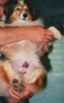
Shetland sheepdog affected with
severe (type 3) vWD
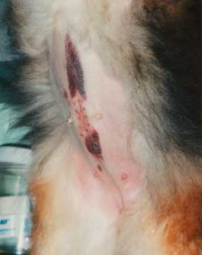
Severe bruising along incision line after
ovariohysterectomy (spay). Bruising occurred in
spite of pre-operative transfusion.
Treatment
Treatment of a severe bleeding episode requires transfusion of canine blood products. Plasma products can be transfused pre-operatively to prevent surgical hemorrhage. Desmopressin acetate (DDAVP) can also be used to improve hemostasis in dogs with mild subtype (Type 1) VWD. Bleeding from minor injuries may be controlled using sutures, bandages or wound glue. Affected dogs should not be given drugs that interfere with normal blood clotting mechanisms. These drugs include aspirin, sulfa-type antibiotics, and heparin.
Laboratory Diagnosis
Laboratory diagnosis of VWD is most often based on results of von Willebrand factor antigen assay (abbreviated VWF:Ag). This test measures the amount or concentration of VWF in a blood sample. The Comparative Coagulation Laboratory reports each dog's result as %VWF:Ag compared to a 100% standard. Dogs having low plasma VWF:Ag (below 50%) are predicted at risk of transmitting or expressing the vWD trait. In general, the most severely affected dogs have marked reduction in plasma VWF:Ag, with values of less than 25%.
The methods used to draw, process, and ship samples are important for accurate VWF:Ag results. Samples containing clots or hemolysis (red cell breakdown) are likely to yield inaccurate or irreproducible results. Use of a standard sampling technique ensures optimum sample quality. See sampling information.
Plasma VWF levels fluctuate somewhat from day to day in normal, healthy dogs. This fluctuation is exaggerated during pregnancy or heat in bitches, and in any dog having a systemic illness (especially liver disease or inflammatory disorders). VWF values are most useful as genetic predictors for VWD status at physiologic “quiet” times, when collected from healthy dogs and bitches not pregnant or in heat. Puppies can be sampled as young as 6 to 8 weeks of age.
Diagnostic ranges of VWF:Ag are used to identify VWD-affected dogs and as an aid for predicting genetic status for the VWD trait.
| Diagnostic Range | vWF:Ag% |
|---|---|
| Normal | 70 to 180 |
| Borderline | 50 to 69 |
| Abnormal | 0 to 49 |
Dogs testing in the normal range are considered clear of the VWD trait, and at low risk for expressing or transmitting vWD.
Dogs testing in the borderline range cannot be accurately classified as carrier or clear on the basis of that measurement. Although at low or no risk of abnormal bleeding, some individuals may be clear and some carriers of the VWD trait. A second VWF:Ag test, screening offspring, and molecular genetic testing can help further clarify a dog’s genetic status.
Dogs testing in the abnormal range are considered carriers of the VWD trait. They are at risk for transmitting an abnormal VWF gene to offspring, and some will express a bleeding tendency. Dogs affected with the most severe subtype (Type 3 VWD) have very low values of VWF:Ag (1% or less).
Inheritance
Hereditary VWD is an autosomal trait, therefore males and females transmit and express the trait with equal frequency. All males and females have 2 VWF genes, one inherited from dam and one from sire. In many breeds, the presence of 1 abnormal VWF gene appears sufficient to cause abnormal and variable bleeding, an expression pattern referred to as "incomplete dominance". Breeds with Type 2 and Type 3 VWD appear to have recessive expression patterns. Clinically affected dogs have 2 abnormal VWF genes, inherited from both dam and sire.
Several distinct VWF gene mutations have been described in different VWD subtypes and affected breeds:
- Type 1 VWD: Splice-site mutation at the end of VWF exon 43 (c.7437G>A)
- Type 2 VWD (German WH and SH Pointers): Missense mutation (c.1657T>G; p.Trp553Gly)
- Type 3 VWD (Scottish terriers): Deletion in VWF exon 4 (c.255Cdel)
- Type 3 VWD (Dutch Kooiker): Splice-site mutation at intron 16 (Int16G>A)
Breeding guidelines based on VWF:Ag diagnostic ranges or genetic tests can help reduce the prevalence of VWD within a family or line, without discriminating against all dogs in that line. Screening for VWD will ensure that no severely affected puppies are produced.
Dogs that test in the normal range (VWF:Ag greater than 70%) are ideal for use in breeding programs. Matings between 2 VDW test-clear parents are predicted to produce only VWD clear pups. Progeny testing (testing parents and entire litter) will further confirm predicted genetic status based on a single VWF:Ag value.
In some cases, dogs that test in the VWD borderline or abnormal range (VWF:Ag < 70%) may be used for breeding, provided they do not express a bleeding tendency. Carriers should be bred to test-clear mates. Some puppies in these matings will test in the normal range. By increasing the number of clear to clear matings in subsequent generations, the proportion of VWD carriers in a line will be gradually reduced, without losing desirable traits. Carrier to carrier matings are undesirable, because these crosses are likely to produce the most severe form of VWD in offspring. Do not breed any dog that expresses abnormal or excessive hemorrhage.
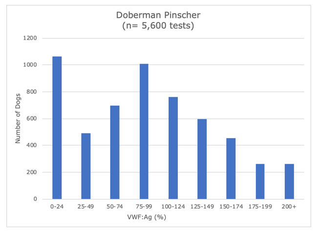
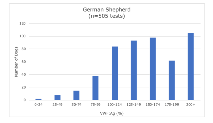
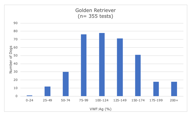
VWD Screening: Breed Summaries
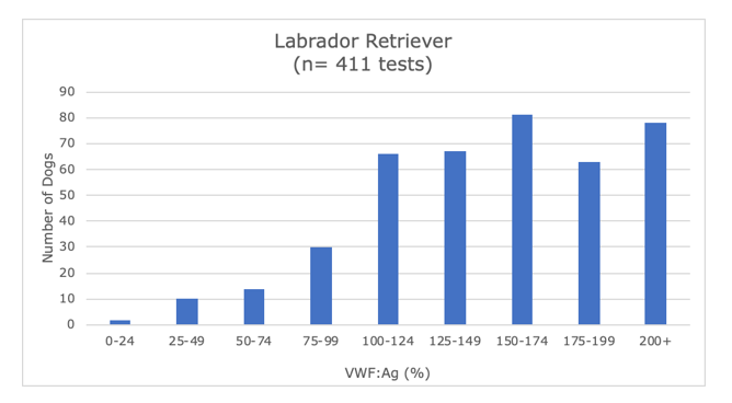
The Comparative Coagulation Laboratory’s submissions for canine VWF:Ag assay were reviewed over a 3-year period (2016 to 2019). The VWF:Ag values for the most commonly tested dogs are summarized by breed in the following graphs:
Doberman Pinscher
Mixed Breed

German shepherd
Golden retriever
Labrador retriever