Schwann cells are named after Theodor Schwann, a German physiologist who discovered these types of cells in the 19th century.
Schwann cell, also called neurilemma cell, are a type of large neurological cell responsible for forming the myelin sheath around the neurons of the peripheral nervous system, and supplying nutrients to individual axons.
Myelin sheath functions to insulate and protect the axons of neurons and is, therefore, important for enhancing the transmission of electrical impulses.
Each Schwann cell comprises a single myelin sheath on an axon. Therefore, numerous Schwann cells are required to myelinate the length of an axon.

Types of Schwann Cells
However, Schwann cells can be either myelinating or non-myelinating. Whilst myelinating Schwann cells wrap around the axons of motor and sensory neurons to form the myelin sheath, non-myelinating Schwann cells do not wrap around the axons but still provide support and cushioning to the unmyelinated axons.
Both types of Schwann cells are vital in the maintenance and regeneration of axons of the neurons within the PNS. Schwann cells originate from the neural crest, which is a group of embryonic cells.
Schwann cells are also considered to be a type of glial cell. Glia cells are non-neuronal cells that do not provide electrical impulses like neurons but function to maintain homeostasis, providing support and protection for neurons.
So, although Schwann cells do not conduct electrical activity themselves, they still help ensure the normal conduction of electrical signals.
Both the myelinating and the non-myelinating Schwann cells are surrounded by sheets of tissue known as basal lamina.
The outside of the basal lamina is covered in a layer of connective tissues known as the endoneurium. The endoneurium contains blood vessels, macrophages, and fibroblasts.
Finally, the inner surface area of the lamina layer faces the plasma membrane of the Schwann cells. Schwann cells could be confused for oligodendrocytes, which are also myelinating cells.
Schwann Cells vs Oligodendrocytes
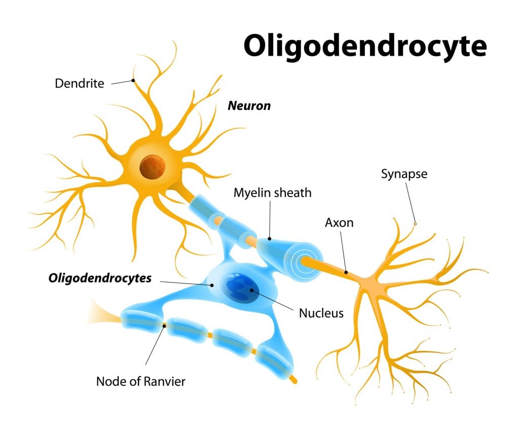
However, whilst Schwann cells myelinate axons of the PNS, the oligodendrocytes provide myelination to axons in the central nervous system (CNS).
Also, each Schwann cell forms a single myelin sheath around an axon, whereas oligodendrocytes form myelin sheaths for multiple surrounding axons. Likewise, Schwann cells are surrounded by basal lamina, whilst the oligodendrocytes are not surrounded by basal.
Schwann Cells Function
Insulation
Myelinating Schwann cells start forming myelin sheaths as early as during the development of the fetus before birth.
For the insulating myelin sheath to be produced by the Schwann cells, the plasma membrane of these cells needs to wrap around the axons of the neuron.
The plasma membrane contains high levels of fat which is essential for constructing the myelin sheath. The plasma membrane works by wrapping itself concentrically around the axon, spiraling to add membrane layers to the myelin sheath.
Sometimes, as many as 100 revolutions of Schwann cell spirals around the neuron’s axon. A Schwann cell that is well-developed may resemble the appearance of a rolled-up sheet of paper, with layers of myelin between each spiral.
A neural-rich protein called Neuregulin is present on the axon’s surface and is necessary for the longevity and maturation of the Schwann cell precursors.
As well as this, the degree of myelination is dependant upon the amount of neuregulin on the axon’s surface. Essentially, the inner layers of the wraps of Schwann cells are typically the membrane, which forms the myelin sheath, whilst the outer layers form the nucleated cytoplasmic layer, called the neurilemma.
It is important that myelin sheath is formed on axons to aid the conduction of electrical signals. Schwann cells that myelinate the axons help to transmit electrical signals at the highest speed so that brain activity is functioning rapidly.
In contrast, the non-myelinating Schwann cells surrounding the axons make them slower conductors of electrical signals. These slow-conducting axons are arranged in a structure that is bundle-like.
Regeneration
In addition to insulating axons, Schwann cells are vital in response to axon damage within the PNS as they can help regenerate the axons.
Any injury to the axon can result in the death of cells and axonal degeneration. When any type of injury occurs, the Schwann cells are sent to the injury site in order to remove dead cells.
The Schwann cells have the capacity to occupy the original space of the neurons and regenerate the fibres in such a way that they are able to return to their original target sites.
The Schwann cells undergo many changes during axonal regeneration. They activate myelin breakdown and up-regulate the expression of cytokines (a large group of proteins secreted by the immune system).
This up-regulation helps to recruit macrophages, which are specialized cells that function to detect and destroy harmful organisms. The macrophages are sent to the site of the injury to clean up the damage.
The Schwann cells increase the number of growth factors, such as neurotrophins, which are proteins that increase the survival and function of neurons. They also secrete proteins such as laminin and collagen and cell adhesion molecules involved in binding with other cells to support the regeneration process.
The stump of a damaged axon can sprout, allowing them to grow. The Schwann cells arrange a regeneration pathway along a tube of the basal lamina. The sprout of the damaged axon can then grow through this tube which helps to stimulate and guide its regeneration.
Due to this, the regenerated axons can reconnect with muscles and organs that they previously controlled with the help of the Schwann cells. Schwann cells are, therefore, especially important as regenerated axons will not reach their target areas without their support.
Damage
Schwann cell dysfunction is primarily associated with demyelinating diseases of the PNS. Demyelination in the PNS describes a pathologic process of destruction of myelin-supporting cells, therefore destroying normal myelin.
These dysfunctions could be a result of genetic mutations, autoimmune responses, infections, and trauma. These causes can impair the myelination process and the functions of the Schwann cells and axons, eventually leading to neurodegeneration.
The insulating myelin segments could be lost or destroyed, and the conduction of neuronal electrical impulses down the axon can be diminished or blocked.
Demyelinating diseases can range from acute to chronic, and some symptoms of these diseases can be characterized as weakened reflexes, weakness, sensory loss, slower nerve conduction, and paralysis.
Disorders that cause damage to the myelin sheath of the PNS, ultimately affecting the function of Schwann cells and axons, are called peripheral demyelinating diseases. Guillain-Barre Syndrome is a rare autoimmune disease affecting the PNS.
With this disorder, the immune system attacks healthy nerve cells in the PNS, resulting in symptoms of weakness and numbness, and may eventually cause paralysis, making it a life-threatening condition if this disease affects the muscles involved with respiration.
Guillain-Barre Syndrome causes damage to the axons of neurons, leading to a blockage of electrical conduction. Although this condition damages the axons, the Schwann cells are also damaged. As a result, producing something called secondary demyelination.
Guillain-Barre Syndrome can be treated through intravenous immunoglobulin, which is a treatment comprised of blood donation that contains healthy antibodies to prevent harmful antibodies from damaging the axons of neurons.
Charcot-Marie-Tooth disease (CMT) is another peripheral demyelinating disorder that is rare and hereditary. CMT occurs when there are mutations in the genes that affect the nerves of the PNS. Both sensory and motor neurons can be affected by this disease, disrupting overall Schwann cell structure and function.
This can affect a lack of growth in Schwann cells as well as abnormal amounts of Schwann cells between the nodes of Ranvier (gaps in the myelin sheath that function to increase the conduction of impulses).
Symptoms of CMT can include having weak muscles, loss of sensation in the feet and legs, difficulty walking, and deformities in the feet, such as having a high arch and bent toes.
CMT usually becomes symptomatic in the feet and legs but may eventually impair functions in the hands and arms. There does not appear to be a cure for CMT. However, patients with this condition can have physical and occupational therapies to help them cope with the symptoms of the disease.
Diabetic neuropathy is another condition that is linked with Schwann cell damage.
This damage can occur with Schwann cells that surround sensory and motor neuron axons. High glucose levels associated with diabetes are a cause of damage to Schwann cells, which can, in turn, increase the risk of neurodegeneration.
Aside from demyelinating diseases, another disease associated with Schwann cell damage includes Schwannomas.
A schwannoma is a tumor that is usually benign but can be harmful and cancerous in rare cases. These tumors grow from the Schwan cells and can occur spontaneously or result from genetic disorders. Although these can be rarely harmful, they can lead to nerve damage and loss of motor control.
In cases where these tumours become large enough to feel under the skin, there may be some pain, as well as muscle weakness, numbness, and tingling sensations.
Interestingly, Schwann cells within the PNS have been implanted into the CNS, specifically the spinal cord of rats, in an attempt to strengthen multiple sclerosis-type damage.
It was found that the Schwann cells formed relatively extensive myelin in the CNS and that blocked nerve impulse conduction improved as a result (Kohama et al., 2001).
Likewise, spinal cord injury has been shown to improve due to Schwann cells. These cells were also implanted into the damaged site in the spinal cord and have been shown to promote axonal regeneration and myelination (Oudega & Xu, 2006).
References
Britannica, T. Editors of Encyclopaedia (2020, July 15). Schwann cell. Encyclopedia Britannica. https://www.britannica.com/science/Schwann-cell
Fallon, M., & Tadi, P. (2019). Histology, Schwann Cells.
Kohama, I., Lankford, K. L., Preiningerova, J., White, F. A., Vollmer, T. L., & Kocsis, J. D. (2001). Transplantation of cryopreserved adult human Schwann cells enhances axonal conduction in demyelinated spinal cord. Journal of Neuroscience, 21 (3), 944-950.
Oudega, M., & Xu, X. M. (2006). Schwann cell transplantation for repair of the adult spinal cord. Journal of neurotrauma, 23 (3-4), 453-467.
Sinha Dutta, S. (2020, February 4). What are Schwann Cells? News Medical Life Sciences. https://www.news-medical.net/health/What-are-Schwann-Cells.aspx#2


