
If you’ve ever wondered how your muscles work or why some muscles are stronger than others, you’re in the right place. In this article, we’ll dive into the exciting world of muscle anatomy, focusing on three key aspects – muscle origin, insertion, and innervation. Whether you’re a bodybuilding enthusiast, a medical student, or just curious about human physiology, understanding these terms will reveal the intricacy behind every move you make. By understanding these terms, you’ll gain a deeper appreciation of your body’s mechanics and how it all works together to create movement. Also, understanding these terms can be extremely beneficial, especially if you’re engaged in bodybuilding or other forms of physical training. You will see how this knowledge can help you optimize your bodybuilding routine.
Knowing your body well is the first step towards optimizing physical potential. Understand the origin, insertion, and innervation of muscles to exercise effectively and prevent injury.
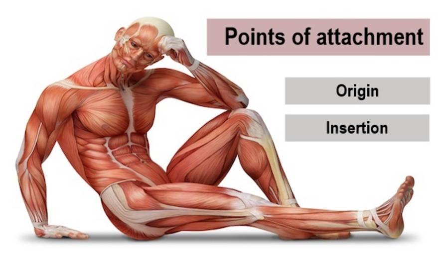
What is muscle attachment?
Of the three types of muscle—smooth, cardiac, and skeletal—it is the third type that attaches to bones, causing them to rotate around joints. It is this combined functioning of muscles, skeletal bones, and joints that allows us to run, lift weights, run on a treadmill, jump, participate in a cycling class, and lift and throw things.
Muscle attachment, simply put, is where a muscle adheres, or attaches, to the body. [1] This attachment typically occurs at both the origin and insertion points of a muscle explained in the next paragraphs.
Interestingly, both the origin and insertion of a muscle are attached to bones, acting as anchor points. The muscle tissue itself doesn’t directly connect to the bone, however. That role goes to a tough, fibrous tissue known as a tendon, which respects the origin and insertion of the muscle.
Tendons are remarkable structures that connect muscle to bone, allowing for the transmission of forces and enabling movement. [1] It’s the tendon that forms the actual attachment to the bone at both the origin and insertion points.
Are muscles only attached to bones? No, it’s not exclusively so. Although muscles commonly attach to bones, they can also attach to other muscles, skin, and various internal organs. This diversity of attachment allows muscles to perform a vast range of movements and functions, from helping us jump or run to basic functions like breathing or moving food through our digestive system.
What is the origin of a muscle?
When we talk about the ‘origin’ of a muscle, we’re zeroing in on where the muscle starts. In general, it’s the attachment point that remains fixed or stable (does not move) during muscle contraction. [2] Also known as the ‘proximal end,’ it’s typically anchored to a larger, more immobile bone like the hip or shoulder. Picture this, it’s the base from which all your muscle fibers reach out and pull or push.
Knowing the origin of a muscle is crucial for a better understanding of how your muscles function. It serves as the foundation, providing the necessary stability for your muscles to generate force. By knowing where the muscle originates, health professionals can predict the muscle’s actions, guide rehabilitation following injury, and even provide therapeutic interventions.
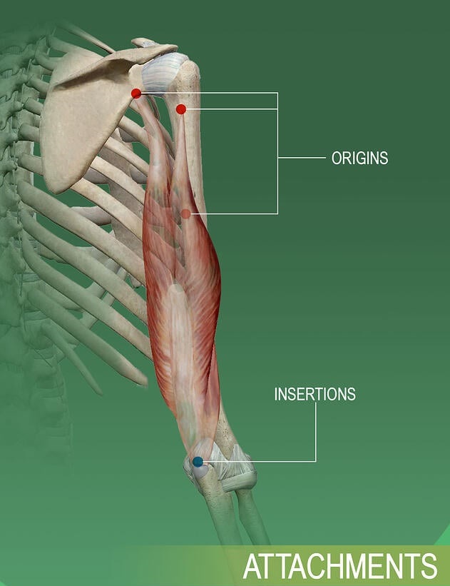
A muscle can have a single origin or several origins known as ‘heads.’ For instance, the triceps brachii muscle in your arm has three origins or ‘heads’ – hence, its name ‘tri-ceps.’ [2] The number of origins a muscle has can influence its strength and range of movement.
Let’s take a look at another example. The biceps brachii in the upper arm has two origin points, hence the prefix ‘bi-‘ in its name. These two points are the long head and the short head, which originate from different points on the scapula and converge to insert on the radius. This dual origin allows for more complex movement and force generation.
What is muscle insertion?
In simple terms, muscle insertion (also known as distal or finish point) refers to the location where a muscle attaches to a bone that typically moves when the muscle contracts. It’s the end of the muscle furthest away from the center of your body. By contracting, the muscle pulls on this bone, leading to movement. [3]
Now, you might be wondering why these points are important, right? Knowing the site of muscle insertion can be crucial because it offers insight into the precise motion your body might undergo when a specific muscle contracts. When your hand curls into a fist, for instance, it’s because your forearm muscles contract and pull on their insertion points in your hand. Understanding this can help delineate the kind of exercises or movements that could specifically target that muscle.
Typically, a muscle has a single insertion point, but it’s possible for a muscle to have multiple insertion points as well. Consider, for example, the deltoid muscle, a large and robust muscle on your shoulder. This muscle has three sections or ‘heads’– anterior, lateral, and posterior– each of which attaches to a different part of your bone structure. In other words, the deltoid muscle has three different insertion points on the humerus. This allows the deltoid to perform a variety of movements, including lifting the arm and rotating the shoulder.
Another great example is your quadriceps, a powerful group of muscles in your thighs. Your quadriceps muscles each have an individual insertion point, but they eventually combine at a common insertion point on your kneecap.
Easy-to-understand examples
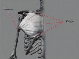
Here is one example for easier understanding. The pectoralis muscle originates on the sternum and inserts on the humerus or upper arm bone. The head that attaches to the arm has the most range of motion, so it is the insertion. The shape of a muscle may change as it moves, but the origin and insertion never change; they are attached to the skeleton.
So by knowing these points you understand how to place the muscle in any pose. You also know what direction the muscle pulls in and what its function is. If you know the start and end points of a muscle, deciphering its function is rather simple. Just remember that muscles pull toward the origin, and you will quickly realize the purpose of the muscle in question. Combine this with useful clues contained in the muscle name, and you have a wealth of information about the form in question.

Biceps brachii is a double-headed muscle, meaning that it has two points of origin or “heads” in the shoulder area. Both heads converge into one muscle, sometimes referred to as the “belly”, that runs the length of the humerus. The short head of each biceps brachii originates at the top of the scapula (at the coracoid process). The long head originates just above the shoulder joint (at the supraglenoid tubercle). Both heads are joined at the elbow (insertion: radial tuberosity).
What is muscle innervation (nerve supply)?
Muscle innervation, also known as nerve supply, pertains to the process by which a nerve interacts with a muscle. Every muscle in your body is connected to a particular nerve or a group of nerves, that controls its movement. These nerves carry messages from your brain to your muscles, instructing them when to contract or relax. It’s these contractions and relaxations that enable you to move your body. So, understanding what muscle innervation is, becomes important. [4]
But why is it important, specifically? Well, knowledge of muscle innervation is crucial in various domains. For healthcare professionals, particularly in fields such as physiotherapy, orthopedics, and sports medicine, understanding muscle innervation helps in diagnosing and treating disorders related to nerves and muscles. For patients recovering from nerve or muscle injuries, understanding how their muscles are innervated is vital to their rehabilitation process. [6]
The significance of muscle innervation isn’t limited to medical contexts. Even if you’re a fitness enthusiast or a bodybuilder, understanding how your muscles are innervated can help you optimize your exercises. By knowing which nerves control which muscles, you can target specific muscles more effectively during your workouts. This not only leads to improved performance and results but also reduces the risk of injuries. So, not only can knowledge of muscle innervation help you in maintaining the health and function of your muscles, but it can also aid in enhancing your physical performance and growth.
Points of origin & insertion and nerve supply (innervation) for the most important muscles in the human body
| Muscle | Origin | Insertion | Innervation |
|---|---|---|---|
| Pectoralis Major – Clavicular Head | Anterior surface of the medial half of the clavicle | Crest of greater tubercle of humerus | Lateral and medial pectoral nerves (C5-C6) |
| Pectoralis Major – Sternal Head | Anterior surface of the sternum & Costal cartilages of the superior six ribs & Aponeurosis of the external oblique | Crest of greater tubercle of humerus | Lateral and medial pectoral nerves (C7-T1) |
| Pectoralis Minor | Anterior surface of ribs 3-5, just lateral to the costal cartilages | Medial border and superior surface of the coracoid process of the scapula | Medial pectoral nerve (C8-T1) |
| Anterior deltoid | The anterior surface of the lateral third of clavicle | Deltoid tuberosity of the lateral humerus | Axillary nerve (C5-C6) |
| Lateral deltoid | Superior surface of acromion process | Deltoid tuberosity of the lateral humerus | Axillary nerve (C5-C6) |
| Posterior deltoid | Inferior border of the spine of the scapula | Deltoid tuberosity of the lateral humerus | Axillary nerve (C5-C6) |
| Supraspinatus Muscle | Supraspinous fossa of the scapula | Greater tubercle of the humerus | Subscapular nerve (C5) |
| Infraspinatus Muscle | Infraspinous fossa of scapula | Middle facet of the greater tubercle of the humerus | Subscapular nerve (C4-C6) |
| Teres Minor Muscle | Middle to upper portion of the lateral border of the scapula, on its posterior surface | Inferior facet of the greater tubercle of the humerus | Axillary nerve (C5-C6) |
| Subscapularis | Subscapular fossa of the scapula | Lesser tubercle of the humerus & Anterior articular capsule of the shoulder joint | Upper and lower subscapular nerve (C5-C6) ok |
| Biceps brachii – Short Head | Tip of the coracoid process of the scapula | Posterior surface of the radial tuberosity & Medial portion of the fascia of the forearm via the bicipital aponeurosis; | Musculocutaneous nerve (C5-C6) |
| Biceps Brachii – Long Head | Supraglenoid tubercle of the scapula | Posterior surface of the radial tuberosity & The medial portion of the fascia of the forearm via the bicipital aponeurosis | Musculocutaneous nerve (C5-C6) |
| Brachialis | Distal half of the anterior surface of the humerus | Coronoid process of the ulna; Tuberosity of ulna | Musculocutaneous nerve (C5-C6) & Radial nerve (C7) |
| Triceps Brachii – Long Head | Infraglenoid tubercle of the scapula | Proximal end of the olecranon of the ulna and fascia of the forearm | Radial nerve (C6-C8) |
| Triceps Brachii – Medial Head | Posterior surface of the humerus (inferior to radial groove) | Proximal end of the olecranon of the ulna and fascia of the forearm | Radial nerve (C6-C8) |
| Triceps Brachii – Lateral Head | The posterior surface of the humerus (superior to radial groove) | Proximal end of the olecranon of the ulna and fascia of the forearm | Radial nerve (C6-C8) |
| Serratus Anterior | External surface of ribs 1-3 (superior fibers); External surface of ribs 4-9 (inferior fibers) | The anterior surface of the medial border of the scapula | Long thoracic nerve (C5-C7) |
| Rectus Abdominis | Pubic symphysis and pubic crest | Xiphoid process and costal cartilages of ribs 5-7 | Thoracoabdominal intercostal nerves (T7-T11) & Subcostal nerve (T12) |
| External Oblique Muscle | External surface of the ribs 5-12 | Anterior iliac crest & Linea alba, inguinal ligament and pubic crest (via external oblique aponeurosis) | Thoracoabdominal intercostal nerves (T7-T11) & Subcostal nerve (T12) & Iliohypogastric nerve (L1) & Ilioinguinal nerve (L1) |
| Internal Oblique | Lumbar fascia & Iliac crest & Inguinal ligament | Linea alba, pubic crest and costal margin (via internal oblique aponeurosis) & Inferior borders of ribs 10-12 | Thoracoabdominal intercostal nerves (T7-T11) & Subcostal nerve (T12) & Iliohypgastric nerve (L1) & Ilioinguinal nerve (L1) |
| Transversus Abdominis | Inguinal ligament & Anterior iliac crest & Lumbar fascia & Costal cartilages of the ribs 7-12 | Linea alba and pubic crest (via transversus abdominis aponeurosis) | Intercostal nerves (T7-T11) & Subcostal nerve (T12) & Iliohypgastric nerve (L1) & Ilioinguinal nerve (L1) |
| Rectus Femoris | Anterior inferior iliac spine & Ilium, superior to the acetabulum | Quadriceps tendon to the base of the patella, and the tibial tuberosity via the patellar ligament & Tibia and knee joint capsule via the medial and lateral patellar retinacula | Femoral nerve (L2-L4) |
| Vastus Lateralis | Greater trochanter and lateral lip of the linea aspera of the femur | Quadriceps tendon to the base of the patella, and the tibial tuberosity via the patellar ligament & Tibia and knee joint capsule via the medial and lateral patellar retinacula | Femoral nerve (L2-L4) |
| Vastus Medialis | Intertrochanteric line and medial lip of the linea aspera of the femur | Quadriceps tendon to the base of the patella, and the tibial tuberosity via the patellar ligament & Tibia and knee joint capsule via the medial and lateral patellar retinacula | Femoral nerve (L2-L4) |
| Vastus Intermedius | Anterior and lateral surfaces of the shaft of the femur | Quadriceps tendon to the base of the patella, and the tibial tuberosity via the patellar ligament & Tibia and knee joint capsule via the medial and lateral patellar retinacula | Femoral nerve (L2-L4) |
| Biceps Femoris Long Head | Ischial tuberosity | Lateral part of the head of the fibula & Lateral collateral ligament & Lateral condyle of the tibia | Tibial division of sciatic nerve part of tibia (L5-S2) |
| Biceps Femoris Short Head | Linea aspera and lateral supracondylar line of the femur | Lateral part of the head of the fibula & Lateral collateral ligament & Lateral condyle of the tibia | Common fibular [peroneal]division of the sciatic nerve (L5-S2) |
| Semitendinosus | Ischial tuberosity | Medial surface of the superior tibia | Tibial division of sciatic nerve part of tibia (L5-S2) |
| Semimembranosus | Ischial tuberosity | Posterior medial condyle of the tibia | Tibial division of sciatic nerve part of tibia (L5-S2) |
| Gluteus Maximus | Dorsal surface of the ilium behind the posterior gluteal line & Posterior surfaces of the sacrum and coccyx & Sacrotuberous ligament | Gluteal tuberosity of femur and iliotibial tract. | Inferior gluteal nerve (L5-S2) |
| Gluteus Medius | Dorsal surface of the ilium between anterior and posterior gluteal lines | Lateral surface of the greater trochanter of the femur | Superior gluteal nerve (L5-S1) |
| Gluteus Minimus | Dorsal surface of the ilium between the anterior gluteal line and inferior gluteal line (just inferior to the gluteus medius origin) | Anterior surface of the greater trochanter of the femur | Superior gluteal nerve (L5-S1) |
| Gastrocnemius | Lateral surface of the lateral condyle of the femur & Popliteal surface of the femur, just superior to the medial condyle of the femur | Posterior surface of the calcaneus via the Achilles tendon & Posterior surface of the calcaneus via the Achilles tendon | Tibial nerve (S1-S2) |
| Soleus | Posterior surface of the head of the fibula & Superior quarter of the posterior surface of the fibula & Soleal line and middle third of medial border of tibia & Tendinous arch of the soleus | Posterior surface of the calcaneus via the Achilles tendon | Tibial nerve (S1-S2) |
| Latissimus Dorsi | Vertebral part: Spinous processes of vertebrae T7-T12, Thoracolumbar fascia Iliac part: Posterior third of crest of ilium Costal part: Ribs 9-12 Scapular part: Inferior angle of scapula | The intertubercular groove of the humerus | Thoracodorsal nerve (C6-C8) |
| Trapezius Muscle | Superior fibers: medial third of the superior nuchal line, external occipital protuberance, nuchal ligament Middle fibers: spinous processes and supraspinous ligaments of vertebrae T1-T4 (or C7-T3) Inferior fibers: spinous processes and supraspinous ligaments of vertebrae T4-T12 | Superior fibers: lateral third of clavicle & Middle fibers: medial acromial margin, superior crest of spine of scapula & Inferior fibers: lateral apex of the medial end of scapular spine | Spinal accessory nerve (CN IX) & Cervical spinal nerves (C3-C4) |
| Rhomboids | Rhomboid Major: Spinous processes of T2-T5 & Rhomboid Minor: Spinous processes of C7-T1 | Rhomboid Major: Medial border of the scapula (from below scapular spine to inferior angle of scapula) & Rhomboid Minor: Medial border of the scapula (in line with scapular spine) | Dorsal scapular nerve (C4-C5) |
The connection between the origin and insertion points of a muscle and its actions
When you understand the relationship between the origin and insertion points of a muscle, you can easily envision how the muscle works. The muscle origin is considered the anchor or start point of the muscle, usually located on the immovable (or less movable) bone, while the muscle insertion refers to the endpoint, attached to the bone that will be moved when the muscle contracts. So, why does this all matter?
- Firstly, the arrangement of the origin and the insertion points determines the trajectory of muscle fiber alignment, directly affecting the range and direction of the movement. For example, when your bicep contracts, the insertion point on your forearm pulls closer to the origin point at the shoulder, thus bending your elbow. [4]
- Secondly, how far apart these two points are is also a factor. It’s a simple principle: the further apart they are, the more potential for movement there is. That’s why your fingers – which have muscles that originate in the elbow – have a vast and versatile range of movement! [8]
- Thirdly, muscles with multiple insertion points – like your deltoid, for example – are capable of producing a higher variety of movements compared to those with a single insertion point. The reason is quite simple. Each insertion point allows the muscle to pull the bone in a different direction. In the case of your deltoid, it has three different insertion points – which coordinate to enable your arm to move upward, forward, and backward. So, yes – multiple insertion points do allow for a greater range of motion and versatility. Again, it’s all about where the muscle anchors and to what point it’s moved towards when it contracts. [7]
To get the bigger picture, here’s a simple rule of thumb: muscles always contract and pull their points of insertion closer to the points of origin. So next time you’re at the gym or even stretching in the morning, take a moment to appreciate the complexity and precision of your body’s design!
Benefits of knowing the exact origin and insertion points of a muscle
Understanding the exact origin and insertion points of a muscle can be enormously beneficial, whether you’re a healthcare provider, an athlete, or merely interested in health and fitness. Here are some key advantages:
- Improved Treatment Planning: For healthcare professionals, knowing the specific origin and insertion points can guide effective treatment strategies, including muscle-related injuries or surgeries.
- Enhanced Athletic Performance: Athletes and fitness enthusiasts can make more informed decisions about their training regimens, understand how particular muscle groups work together, and improve their overall performance.
- Precise Movement and Exercise: A comprehensive understanding of muscle origin and insertion points can assist you in performing exercises with accurate form and movement. This can reduce the risk of strain and injury and ensure that the targeted muscle groups are acted upon efficiently.
- Bodybuilding and Physical Development: For those engaged in bodybuilding, a precise knowledge of muscle origin and insertion points can help in the effective isolation and targeting of specific muscles for the most desired growth and symmetry.
- Pain Management: If you’re dealing with chronic muscle pain or muscular disorders, this knowledge can help you better understand the nature of your discomfort and guide physical therapy efforts to mitigate such issues.
- Better Self-Knowledge: Finally, understanding muscle origins and insertion points can contribute considerably to your self-knowledge. In terms of health, wellness, and personal fitness, education is power, and understanding your body can lead to more informed lifestyle choices.
How can I apply my knowledge of muscle anatomy to my bodybuilding routine?
When you refine your understanding of muscle anatomy, particularly muscle origins, insertions, and innervation, your approach to bodybuilding can dramatically evolve. But how, you might ask? Let’s break it down.
First, knowing the origin and insertion points of a muscle helps you target your workouts more effectively. For instance, understanding that the biceps brachii originates from the shoulder blade and inserts into the forearm can help you design exercise regimes that target this muscle directly and maximize its development.
Next, a muscle can have multiple origins. For example, the trapezius muscle, a large muscle in your back, has multiple origins along the spine and inserts on the shoulder blades and back of the neck. Knowing these points helps you identify the extent of the muscle and ensures that exercises fully engage the whole muscle, rather than just a portion of it.
Multiple insertion points are also possible, though less common. A muscle with multiple insertions, like the deltoid muscle, has fibers that all converge on a single tendon. To train such a muscle, you would need a variety of exercises that target all fibers and insertion points, increasing its overall shape and size. That’s why we have special exercises for each head of the deltoid muscle. Therefore, to fully hit your delts you must pick at least one exercise for each deltoid head.
Finally, the innervation (or nerve supply) to your muscles is crucial. During workouts, nerves supply information from your brain to your muscles, triggering the muscle contraction necessary for movement. If you know a muscle’s innervation, you can work towards maximizing muscle stimulation and growth. For instance, knowing that the radial nerve innervates the triceps brachii can help you utilize exercises that fully engage this nerve, leading to better contraction and muscle development.
Muscle insertions – importance in professional bodybuilding
Muscle insertions determine to what degree a specific muscle can be developed. The easiest examples to illustrate this are the biceps and the calves. Your biceps are either short, average, or long. You can figure this out in two seconds by flexing your bicep with the forearm and upper arm at a right angle. How much space is there between the end of your biceps and your elbow?
If there’s a lot of room there, your biceps are short. In case there’s about an inch of space, consider your muscle length average. On the other hand, if there’s no room at all and your biceps muscle is jammed right up against the elbow joint, and in that case you have long biceps. If a muscle is attached lower on the bone, that muscle will look longer when fully developed and will create a more pleasing appearance.
Having long muscles means that you have more muscle cells to work with and increase the size.
The calves are the same way, with the two extremes being high and low calves. Flex your calf and see where the muscle ends on its way to your ankle. If it stops not too far down from your knee, you are unfortunately stuck with high calves. You can beat the crap out of them but they will always be a weak point on your body. If they go way down you are in luck. You probably have nice calves without even training them.
Every muscle on your body inserts wherever it has been programmed to end up by your particular DNA. All this nonsense about preacher curls being able to “fill in” short biceps is nothing but wishful thinking.
Summary
A skeletal muscle usually connects to the bone, or occasionally other muscles or tissues, at a minimum of two different points. If it is attached to a bone that remains static during a certain movement, this attachment is referred to as the origin. On the other hand, if it is attached to a bone that moves during a particular action, this attachment is known as the insertion.
Muscle innervation refers to the supply of nerves to a muscle, enabling it to respond to the brain’s commands for movement. In essence, it’s the process that allows nerves to connect with and control muscle activity.
Understanding these terms and concepts is crucial in the field of bodybuilding. Knowing the origin and insertion points of a muscle can help in targeting specific muscles during workouts, potentially leading to more effective training. Similarly, understanding muscle innervation can provide insight into the body’s response to exercise and the development of muscle strength and coordination.