

In this explainer, we will learn how to describe the structure and function of membranes and describe how factors affecting membrane permeability can be investigated.
Every living organism, whether eukaryotic/prokaryotic or animal or bacterium, possesses cells that are surrounded by a membrane. These membranes act as the gatekeepers of the cell, controlling what enters and leaves, among several other important roles.
Eukaryotic cells even have membranes within the cells themselves! These intracellular membranes are found surrounding eukaryotic organelles; they allow different conditions to be present and therefore different chemical reactions to occur in each separate region of the cell. This separation of the cell components is called compartmentalization. Compartmentalization of organelles in a cell can be likened to how rooms in a house are separated from each other.
Definition: Compartmentalization
Compartmentalization is the formation of separate membrane-bound areas within a cell to allow incompatible reactions to occur within different parts of the cell.
An organelle is a subcellular structure that carries out a specific function. It may or may not be compartmentalized by a membrane.
Definition: Organelle
An organelle is a subcellular structure that carries out a specific function.
A membrane that surrounds an organelle may be a single layer, or two layers, referred to as a double membrane. Organelles that have double membranes include the nucleus, mitochondria, and chloroplasts. Organelles that have a single membrane include lysosomes and the Golgi apparatus, sometimes called the Golgi body.
The eukaryotic intracellular organelle membranes are also often a site of chemical reactions. The rough endoplasmic reticulum (RER) is one such example, with ribosomes embedded in its membrane to carry out protein synthesis. This makes the RER appear “rough” in contrast to the smooth endoplasmic reticulum (SER), which has no ribosomes. Both RER and SER have a single membrane. The inner membrane of mitochondria and membranes of chloroplasts are the sites of aerobic respiration and some reactions in photosynthesis respectively.
Example 1: Identifying Organelles with Membranes and Describing Their Function
Eukaryotic cells contain many organelles with internal membranes.
- Which of the following organelles does not have membranes as part of its structure?
- The ribosome
- The Golgi apparatus
- The endoplasmic reticulum
- The nucleus
- The lysosome
- Internal membranes provide areas with different conditions for specific reactions to take place within a cell.
What term describes this structural adaptation of eukaryotic cells?
Answer
Part 1
In addition to membranes surrounding a cell, many organelles within eukaryotic cells also have membranes. An organelle is a subcellular structure that carries out a specific function.
Depending on the organelle, it may be surrounded by one membrane, two membranes, or no membrane at all. For example, ribosomes and centrosomes are organelles that are not surrounded by a membrane, while lysosomes, the endoplasmic reticulum, and the Golgi apparatus have a single membrane and the nucleus and mitochondria each have a double membrane!
Therefore, the organelle in the list that does not have a membrane as part of its structure is the ribosome.
Part 2
The membranes that surround some organelles separate their contents from their surroundings and allow different conditions to be present within each membrane-bound organelle. This means that different chemical reactions can occur throughout the cell within the organelles, but reactions can also occur across the membranes themselves.
One example is the rough endoplasmic reticulum (RER). The RER has ribosomes on its surface that carry out protein synthesis but do not have membranes. The separation of the contents of an organelle from its external environment by a membrane is called compartmentalization.
Therefore, the term that describes the structural adaptation of internal membranes in eukaryotic cells is compartmentalization.
Depending on their location and composition, membranes have a whole range of functions, but the key point common to them all is that they act to separate a cell, or different sections of a cell, from their surroundings.
Cellular membranes are often described as semipermeable, meaning that only certain molecules can pass through them, using proteins embedded in their surface. The membranes can be likened to walls separating the rooms in a house. They control which substances will move into and out of the cell, with proteins acting effectively as the “doors” to each “room” which can be “locked” or “unlocked” for specific substances.
Whether they are internal cell membranes or are at the cell surface, all cellular membranes have the same basic structure which we will look at first, before learning their functions and the factors that affect their permeability.
Definition: Plasma Membrane
A plasma membrane is a semipermeable barrier consisting of a bilayer of phospholipids and other embedded molecules. Plasma membranes surround all cells, separating them from their external environment.
All membranes, for example, the cell surface membrane of a typical animal cell in Figure 1, consist of a structure called the phospholipid bilayer.

Let’s break down the term phospholipid bilayer to understand it better: membranes are mostly composed of molecules called phospholipids, which have a hydrophilic (water-loving) phosphate head and a hydrophobic (water-fearing) fatty acid tail.
The word bilayer refers to the fact that there are two layers of these phospholipids, one facing outward to the external environment and one facing inward to the contents of the cell cytoplasm (see Figure 1). Hydrophilic phosphate heads are attracted to water molecules.
As most cells, and components within them, are in an aqueous environment (containing water), the hydrophilic phosphate heads arrange themselves to be close to them on both the internal and external surfaces of the membrane. The fatty acid tails however are repelled by the water molecules and so arrange themselves facing inward and toward each other as seen in Figure 1.
It is useful to note that the hydrophobic fatty acid tails in the phospholipid bilayer can consist of fatty acids that are either saturated or unsaturated. You can see a saturated and an unsaturated fatty acid in the tail of the phospholipid molecule in Figure 2 below.

You might have noticed that the structures of the saturated and unsaturated fatty acids that make up the tail of this phospholipid molecule differ. Saturated fatty acids consist of single bonds between all carbon atoms, forming a straight chain of carbons that are completely saturated with hydrogen atoms. Unsaturated fatty acids contain at least one double bond between the two carbons at the point of the “kink” in the tail, so it is not fully saturated with hydrogen atoms.
These kinks mean that adjacent fatty acid tails cannot pack as tightly together compared to tails made of saturated fatty acids, which increases the fluidity of the plasma membrane.
Definition: Phospholipid Bilayer
The phospholipid bilayer is a membrane consisting of two layers of lipid molecules with a hydrophilic phosphate head and hydrophobic fatty acid tails.
Key Term: Hydrophilic Phosphate Heads
Hydrophilic phosphate heads are the parts of phospholipid molecules that are attracted to water and therefore form the surface of cellular membranes that are in aqueous environments.
Key Term: Hydrophobic Fatty Acid Tails
Hydrophobic fatty acid tails are the parts of phospholipid molecules that are repelled by water and therefore form the inner core of cellular membranes.
Example 2: Describing the Structure of Biological Membranes
Which of the following statements about the structure of biological membranes is correct?
- Cell membranes are formed from two layers of phospholipids, where the hydrophilic phospholipid heads face outward.
- Cell membranes are formed from a single layer of phospholipids, connected by their hydrophobic heads.
- Cell membranes are formed from two layers of phospholipids, where the hydrophilic phospholipid tails link together in a matrix.
- Cell membranes are formed from a single layer of phospholipids and a single layer of glycolipids, connected by hydrophobic tails.
Answer
Cell membranes are described as a phospholipid bilayer. This means that it consists of two layers of phospholipid molecules adjacent to each other. Cells are usually in an aqueous environment, containing water molecules, and the cell cytoplasm is also aqueous. This means that the hydrophilic (water-loving) heads of the phospholipid molecules face outward to associate with the water molecules in the exterior and interior environments of the cell. The fatty acid tails of the phospholipid molecules are hydrophobic (water fearing) and so are repelled by the water molecules in the cytoplasm and outside the cell and face inward toward each other.

Therefore, the correct statement describing the structure of the cell surface membrane is A: cell membranes are formed from two layers of phospholipids, where the hydrophilic phospholipid heads face outward.
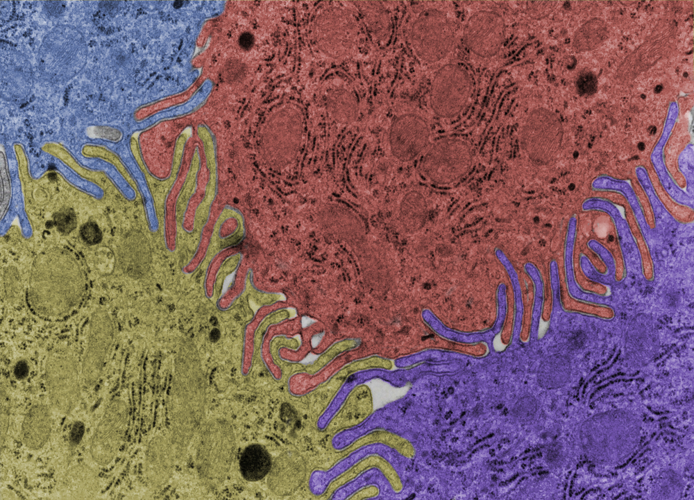
When it was first observed under an electron microscope, the phospholipid bilayer appeared as “tram tracks,” which helped scientists determine that the membrane had two layers. You might be able to see why they were called “tram tracks” by looking at the false-colored micrograph showing the phospholipid bilayers at the border of four different epithelial cells below.
This tram track observation helped scientists develop the fluid mosaic model, describing how the phospholipids are, to a certain degree, free to move fluidly throughout the membrane as they are not chemically bound to each other but simply attracted to or repelled by water molecules.
The term mosaic refers to the molecules (mostly proteins) of different shapes and sizes that are embedded in various sections of the plasma membrane, much like tiles of a mosaic. We are now going to discuss the roles of these different “mosaic” molecules that are visible in Figure 4 below.
Key Term: Fluid Mosaic Model
The fluid mosaic model is a way of understanding how cellular membranes function as the phospholipids are fluid, moving freely within the membrane with molecules of different shapes and sizes embedded in the membrane like mosaic tiles.

There are various proteins that embed themselves in the phospholipid bilayer. One type is called an intrinsic protein (transmembrane or integral protein), which spans both layers of the membrane as in Figure 4. All intrinsic proteins are globular proteins with hydrophobic regions that fit into the membrane.
The example in Figure 4 is a channel protein. These contain a hydrophilic pore through which polar molecules and ions can travel passively by diffusion down the concentration gradient. These substances would otherwise not be able to be transported across the membrane due to the hydrophobic nature of the phospholipid fatty acid tails.
Carrier proteins are another type of intrinsic protein, modeled in Figure 5. They contain receptors specific to one molecule.
For example, in Figure 5, glucose is being transported from the extracellular space into the cell. Due to the specific shape of the receptor, no other molecules would fit into the glucose carrier protein. It may help to visualize glucose as the only molecule that has a “key” for this specific protein “door.”
When glucose binds to the receptor on the carrier protein, the protein changes shape and releases glucose on the other side of the membrane. Depending on the concentration of glucose inside and outside the cell, it may need to be either actively or passively transported by carrier proteins.
Active transport requires energy to transport molecules because they are moving against their concentration gradient. Passive transport does not require energy to transport molecules as they are moving down their concentration gradient. If there was a high concentration of glucose outside the cell and a lower concentration inside, for example, glucose would move into the cell down its concentration gradient by passive transport, using the carrier proteins but no energy.

Key Term: Intrinsic Proteins
Intrinsic proteins are embedded through both layers of a membrane and can be carrier proteins for transporting substances actively or passively into or out of the cell, channel proteins for passive transport only, or others.
Key Term: Channel Proteins
Channel proteins are intrinsic proteins that provide a hydrophilic channel allowing the diffusion of polar molecules and ions down their concentration gradient through membranes.
Key Term: Carrier Proteins
Carrier proteins are intrinsic proteins that have a receptor with a specific shape complementary to certain molecules, and upon binding to them, they undergo a change in shape to transport the molecules through a membrane either up (active transport) or down (passive transport) their concentration gradient.
Glycoproteins are an example of an intrinsic protein, also visible in Figure 4. The glyco- prefix of the word indicates that the protein is attached to a carbohydrate (sugar) chain. Glycoproteins help cells adhere to each other and stick together, and they can also act as receptors.
The general role of a receptor is to allow a specific chemical, such as a hormone, to bind to it and to elicit a response in the cell. This is called cell signaling and it allows the cells of the body to communicate with each other efficiently.
Key Term: Glycoproteins
Glycoproteins are a type of intrinsic protein attached to a carbohydrate chain that help cells adhere to each other and act as receptors for different chemicals to elicit a response in the cell (cell signaling).
Glycolipids, also visible in Figure 4, are like glycoproteins, but instead of a carbohydrate chain attaching to an intrinsic protein, they attach directly to the phospholipids in the bilayer. They are sometimes referred to as cell markers or antigens, as they are used by the immune system in cell recognition to identify cells as self or nonself. If cells are nonself, it indicates they do not belong to the organism itself and are therefore possibly harmful and must be destroyed.
Definition: Glycolipids
Glycolipids are lipids attached to carbohydrate chains, often called cell markers or antigens, that can be recognized by the immune system as self or nonself, the latter indicating they are attached to cells from another organism such as a pathogen.
An example of glycolipids in human cells is in ABO blood grouping. They are glycolipids on the cell surface membrane of red blood cells that act as antigens, specific for one of four blood groups: A, B, AB, and O.
Extrinsic proteins, or peripheral proteins, do not cross the entire diameter of the membrane like intrinsic proteins as can be seen in Figure 4. Instead, they are embedded on just one side of the phospholipid bilayer, interacting with the phosphate head of a phospholipid on either the interior or exterior surface of the membrane or with an intrinsic protein.
Definition: Extrinsic Proteins
Extrinsic proteins are peripheral proteins embedded only on one side of the membrane, interacting with either the phosphate head of a phospholipid or with embedded intrinsic proteins.
The final structure we will discuss is cholesterol, visible in Figure 4 as a small red structure embedded between phospholipid molecules. Cholesterol is an abundant lipid in the membrane of animal cells. It has a hydrophilic head and a rigid, bulky hydrophobic tail that interacts with the phospholipid tails in the membrane. Through these interactions that modulate the packing of phospholipids in the membrane, cholesterol can either increase or reduce the fluidity of membranes in the function of temperature as we will see now.
Key Term: Cholesterol
Cholesterol is a lipid molecule that regulates the fluidity of membranes over a wide range of temperatures by fitting between the phospholipid molecules.
Example 3: Describing the Movement of Molecules through the Plasma Membrane
Hydrogen cyanide is a toxic small nonpolar molecule that is released by some plants to deter herbivores. Cyanide crosses membranes and inhibits a key process in respiration.

What is the most likely way for this molecule to pass through membranes?
- It diffuses through the phospholipid bilayer.
- It diffuses in the cell using a carrier protein.
- It enters through a channel protein.
- It enters by endocytosis.
Answer
When deducing the method by which a molecule is passing through a plasma membrane, look at the information given about the molecule in the question. We can see that hydrogen cyanide is small and nonpolar.
Endocytosis is an example of bulk transport by which large quantities of molecules are actively transported into a cell using a vesicle (endo- meaning into, cyto- meaning cell). is unlikely to be in such large quantities that endocytosis will be required. For this particular question, we can assume that the concentration of inside the herbivore’s cells is much lower than outside as the molecule is toxic by inhibiting one of the processes in respiration. This would imply that passive transport is occurring, but as all of our options could be passive aside from endocytosis, this does not help us narrow down our options much beyond eliminating endocytosis.
Large or polar molecules are unable to diffuse directly across membranes and so must use either a carrier or channel protein. This molecule is stated to be both small and nonpolar, suggesting that it does not require a protein and can simply diffuse across the phospholipids.
Our correct answer is, therefore, A: it diffuses through the phospholipid bilayer.
Temperature has a significant impact on the fluidity of cellular membranes.
At low temperatures, phospholipids tend to move less and become more tightly organized in a crystal-like structure. Therefore, low temperatures make membranes more rigid which can interfere with key functions like the passage of gases. More rigidity also makes membranes more prone to break. Cholesterol limits this phenomenon by disrupting the phospholipids’ tight and regular organization with its bulky tail, thereby increasing membrane fluidity.
At higher temperatures, more thermal (heat) energy is supplied to the phospholipids in the bilayer, which increases their kinetic energy, or movement speed, increasing the fluidity of the membrane. If the membrane is more fluid, its permeability increases as there are many more spaces through which substances can move. Another issue is that the embedded proteins can become denatured (irreversibly changing shape) if the temperature rises above their optimum level. This means that they can no longer carry out their transport roles.
At higher temperature, however, the cholesterol tail keeps strong interactions with the phospholipids around and pulls them together, which prevents the membrane from becoming too fluid. In conclusion, cholesterol acts like a buffer that maintains a stable membrane fluidity over a wide range of temperatures.
Organic solvents that are less polar than water, such as alcohols or nonpolar solvents, are capable of dissolving plasma membranes. An example of this is in antiseptic: the alcohol disrupts the membranes of bacterial cells by dissolving the fatty acids of the phospholipids, killing the bacteria. Alcohol is present in alcoholic drinks, which disorders cell membranes of neurons. This disrupts the transmission of nerve impulses and makes the person’s reactions slower, among other behavioral side effects. Some more hydrophobic substances, such as certain drugs, are also able to pass the membrane directly.
Example 4: Describing Conditions That Damage Cell Membranes
Which of the following conditions is most likely to damage a cell membrane and increase its permeability?
- Low temperatures (4–)
- Hypertonic solutions
- Organic solvents
- Polar solvents
Answer
High temperatures increase the permeability of a cell surface membrane by increasing the kinetic energy and therefore fluidity of the phospholipids in the membrane, making the membrane more “leaky.” Very high temperatures can also increase permeability by irreversibly denaturing proteins involved in transport of substances across the membrane, so they can no longer function effectively.
Organic solvents also increase permeability of a cell surface membrane by dissolving the phospholipids in the membrane. This is true for all solvents that are less polar than water, as they cannot interact with the phospholipids in the bilayer in the same manner as polar water molecules would, so they cannot maintain the bilayer shape.
Hypertonic (hyper-, so you know it has a high solute concentration) or hypotonic (hypo-, so you know it has a low solute concentration) solutions may impact the rate of movement of molecules of solute and/or water across the plasma membrane, but this will not be likely to drastically affect membrane permeability.
Therefore, the correct answer describing the likely effects of different conditions on increasing the permeability of the cell surface membrane is C: organic solvents.
There are some experiments that can be conducted to investigate the effects of temperature and organic solvent concentration on membrane structure and permeability. Such experiments usually involve a process called colorimetry, which is a technique used to measure the concentration of colored compounds or pigments in a solution.
Colorimetry is especially useful when the experimental cells contain pigment that is highly concentrated. Beetroot cells contain the red pigment betalain that gives them a distinctive dark red color, and they are often used in colorimetry. Figure 6 below shows an example of how colorimetry works.

Key Term: Colorimeter
A colorimeter compares the amount of light passing through a sample solution with the amount of light that can pass through a sample of pure solvent that is entirely transparent.
Key Term: Light Absorbance
Light absorbance is a measure of the quantity of light absorbed by a sample solution in a colorimeter.
When placed into a solution of water, some of the red pigment will leak out of the beetroot cells through their cell surface membranes into the solution. The more permeable the cell surface membrane is, the more the pigment that will leak out. The solution is then placed into a square vial called a cuvette, which is placed in a colorimeter, seen as the red sample in Figure 6.
The colorimeter is a device that passes light through a solution and measures the percentage of light that has been transmitted through and the percentage absorbed by the pigment. If all light passes through a sample, none was absorbed by the pigment, so the absorbance would be zero and the transmittance to the detector seen in Figure 6 would be .
Specific color filters are usually selected based on the color of the sample of study and placed between the light source and the sample to give more accurate values of absorbance by the pigment.
The graph in Figure 7 below displays some sample results from this experiment. You can see that as the temperature increases, the absorbance of blue light by the red betalain pigment increases.

Let’s recap some of the key points we have covered in this explainer.
Key Points
- Plasma membranes surround every living cell, and eukaryotes also have intracellular membranes surrounding some of their organelles for compartmentalization.
- The fluid mosaic model describes how the cellular membrane consists of freely moving phospholipids with hydrophilic phosphate heads and hydrophobic fatty acid tails, interspersed with embedded proteins and cholesterol.
- High temperature affects membrane permeability by increasing the fluidity of the phospholipids and denaturing embedded proteins.
- Organic solvents, such as alcohols, also affect membrane permeability by dissolving the phospholipids making the membrane more fluid and permeable.
- Membrane permeability can be investigated using a colorimeter to measure absorbance of light through a solution into which pigment has leaked through a membrane.

