


INTRODUCTION
From early childhood to later stages of life, many dental patients experience a variety of treatments, ranging from basic fillings for cavities, to more complex procedures like crowns and bridges. Throughout their lives, they may also require specialized care from endodontists, orthodontists, or periodontists to maintain their oral health as they age. With an average lifespan of 77.5 years for both sexes,1 and the percentage of older adults retaining their natural teeth on the rise,2 the dental profession in the United States is seeing an older patient population presenting for treatment who have retained most, if not all, of their natural dentition.
However, as one of the most mobile countries in the world,3 these patients have been treated by a variety of dental professionals as they relocate from one locale to the next. Whether moving to upsize or downsize their homes, change jobs, or retire to a warmer climate, it is estimated that the average American moves 11.7 times in his or her lifetime.4 Moving to a larger city or across state lines necessitates finding a new dentist to meet his or her prevention and treatment needs. Other issues impacting the move to a new practice may be dissatisfaction with treatment results, the retirement of the primary care dentist, or high out-of-pocket expenses. For older adults in particular, the result can be a patchwork of dentistry performed to address immediate needs, which, over a lifetime, have lost the continuity and history of previous treatment goals.
In the case presented here, an older patient in good health presented to the practice with aesthetic concerns and had all but given up on addressing her primary complaint of jaw pain after a variety of treatment modalities meant to treat it had failed.
CASE REPORT
A 68-year-old female was referred to the practice by her husband (Figure 1). At her initial consultation, the patient recounted her dental history of orthodontic and prosthodontic treatment to relieve her TMJ pain and address aesthetic concerns. Her TMJ issues had plagued her since the age of 27 and still persisted, making her skeptical that any treatment would be successful. She was also concerned about the chipping occurring on her 2 natural central incisors (Figure 2).

Figure 1. Preoperative photo of the patient revealing her gummy smile and lack of gingival harmony.

Figure 2. Close-up view of the anterior teeth revealed chipping on the central incisors.
A series of x-rays were taken to ensure the structural soundness and health of the patient’s teeth, gums, and bone. Preoperative photos of the maxillary arch documented that teeth Nos. 4 and 5 had been restored with crowns; No. 7 was a crown over a 30-year-old titanium implant; and Nos. 2, 3, 14, and 15 had undergone root canal treatment (Figure 3). Kois glasses also revealed a slight midline discrepancy, which would also need to be addressed (Figure 4).

Figure 3. X-rays and pre-op photos of the maxillary arch documented that teeth Nos. 4 and 5 had been restored with crowns; No. 7 was a crown over a 30-year-old titanium implant; and teeth Nos. 2, 3, 14, and 15 had undergone root canal treatment.

Figure 4. Kois glasses revealed her midline alignment would need to be addressed.
On the mandibular arch teeth, Nos. 23 to 26 had been treated with veneers, No. 19 was a crown, Nos. 20 to 22 and 27 to 29 were porcelain bridges, and natural teeth Nos. 30 and 31 were both restored with large composite fillings (Figure 5). Severe pitting on tooth No. 30 along the buccal cusp was noted, along with wear on the cusps of the other mandibular molars and crack lines on her 2 anterior central incisors (Figure 6). The patient exhibited a VDO of only 32 mm with evidence of a collapsed bite. She had a wide gummy smile (Figure 7) with poor gingival symmetry of the anterior arch.

Figure 5. Pre-op view of the mandibular arch teeth showed that teeth Nos. 23 to 26 had been treated with veneers, No. 19 was a crown, Nos. 20 to 22 and 27 to 29 were porcelain bridges, and natural teeth Nos. 30 and 31 had both been restored with large composite fillings.

Figure 6. Although not an immediate problem, crack lines in the central incisors could cause issues in the future.

Figure 7. The patient exhibited a VDO of only 32 mm with evidence of a collapsed bite, a wide gummy smile, and poor gingival symmetry.
Treatment Proposal
In order to restore proper function and aesthetics for this patient, the first step in the treatment plan was to address her TMJ pain. To determine if a Kois Deprogrammer would be a viable treatment, load (joint) and immobility (muscle) tests were conducted and were deemed negative, indicating a Kois device would be an appropriate treatment strategy.5-9 A Kois Deprogrammer was prescribed to restore her occlusal and temporomandibular stability and open her VDO to create restorative space for a full-mouth rehabilitation. The goal was to open her bite 1.5 mm in the posterior and restore her upper arch with crowns on teeth Nos. 2 to 15 and then, at a later appointment, restore her lower arch. A leaf gauge was used to determine centric relation. Intraoral scans of both arches (Figure 8), a bite registration, and pre-op photos were sent to the laboratory for fabrication of a Kois Deprogrammer. At the second appointment, the deprogramming device was fitted, and the patient was instructed to wear it 24 hours per day for 2 weeks except when eating.
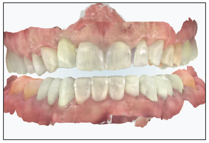
Figure 8. After testing load and mobility tests of her jaw, a Kois Deprogrammer was prescribed to relieve her TMJ pain. Intraoral scans of both arches, a bite registration, and pre-op photos were sent to the laboratory for fabrication of a Kois Deprogrammer.
Two weeks later, the patient reported that she was free from pain and that she could now open to 45 mm. Scans of both arches of the deprogrammed jaw and a bite relation photo (Figure 9) were taken and sent to the laboratory for fabrication of a diagnostic wax-up and a putty matrix for provisionalization.

Figure 9. Post-deprogramming scans of both arches and a bite relation photo were sent to the laboratory for fabrication of a diagnostic wax-up and a putty matrix for provisionalization.
Restorative Treatment
The patient’s excessive gum display was addressed on the fourth appointment in order to achieve the desired aesthetic outcome.10 In this case, gum recession on the implant tooth No. 7 guided the crown-lengthening surgery performed on teeth Nos. 4 to 13. All paper records of the 30-year-old implant had been purged, leaving no option but to prep it for a crown with no knowledge of the implant system used and to mask the metallic substructure as best as possible.

Figures 10 to 12. Crown-lengthening surgery for this case was guided by the gum recession on implant tooth No. 7. After 6 weeks, the gums had healed nicely.
When the patient returned 6 weeks later, the gums had healed nicely (Figures 10 to 12). The crowns on teeth Nos. 2 to 5, 7, 14, and 15 were removed and examined for underlying decay, and all teeth in the upper arch were prepared to receive all-ceramic crowns (Figure 13). Photos of the prepared patient with the stump shade were taken (Figure 14) for communication with the laboratory.

Figure 13. The upper arch was prepared for all-ceramic pressed crowns on teeth Nos. 2 to 15.

Figure 14. A photo of the stump shade was taken to communicate with the laboratory. The key to restorative success would be masking the titanium abutment on tooth No. 7.

Figures 15 to 17. The upper arch was provisionalized, and the patient was instructed to wear the provisionals for a month to test fit, function, and aesthetics.
Using the putty matrix supplied by the laboratory, provisionals (Structur 2 SC Self Cure [VOCO]) were fabricated for both arches chairside and spot-etched and bonded in place (Adhese [Ivoclar]) (Figures 15 to 17). For the mandibular arch, all existing restorations were removed, and the teeth were provisionalized without prepping to ensure the patient functioned well with her new centric relation. The patient approved the aesthetics of the provisionals and was advised to wear them for one month to test the function and fit as well as to ensure her jaw pain was no longer an issue.
After wearing the provisionals for a month, she returned to the practice pleased with the aesthetics and functional outcome of the treatment and reported no jaw pain was present. A scan of the upper arch, along with photos with a tooth shade tab (Figure 18) and a bite stick photo were sent to the laboratory to fabricate the final restorations (IPS e.max Press shade M1 [Ivoclar]).

Figure 18. A photo of the provisionals with a shade tab was taken to communicate with the laboratory.

Figures 19 to 22. The final maxillary restorations were delivered and seated.
On delivery, the final restorations for the maxillary arch were seated (Variolink Esthetic DC Neutral [Ivoclar]) (Figures 19 to 22). The following day, the teeth in the mandibular arch were prepared and provisionalized. Upon delivery, the final mandibular restorations were seated (Variolink Esthetic DC for crowns and Variolink Esthetic LC Neutral for veneers [Ivoclar]), and the patient was pleased with the final outcome. A month later, she returned for a follow-up appointment, and final photos were taken of the case outcome (Figure 23).

Figure 23. Three weeks after the final mandibular restorations were seated, the patient returned to the practice for final photographs.
CONCLUSION
Often, patients present to the practice disappointed by past dental work and have lost faith and trust in the profession in treating long-standing problems. Taking them to the point of accepting treatment involves rebuilding that trust and helping them overcome their fear of the process. Approaching these patients with concern and compassion and focusing on relieving long-standing issues before rehabilitating the aesthetics of their smiles can restore that trust and belief in the profession.
The patient’s reaction sums up the outcome of this case: “No doubt my case brought with it obstacles and challenges. As a patient, coming to the decision of going forward with a full restoration is actually a big decision. Turning oneself over to the process involves trust and letting go of fear. I came to you to change, improve, and restore my smile. You did that and far more by providing me with a professional, all-encompassing plan of action to correct and restore long-standing problems. I remain grateful and appreciative. Thank you kindly.”
ACKOWLEDGMENTS
Dr. Desai would like to thank ceramist, Juan Rego, from Smile Designs by Rego (regosmiles.com) for the collaboration and ceramic work.
REFERENCES
1. Kochanek KD, Murphy SL, Xu J, et al. Mortality in the United States, 2022. NCHS Data Brief. 2022;(492):1-8.
2. Centers for Disease Control and Prevention. Oral Health Surveillance Report: Trends in Dental Caries and Sealants, Tooth Retention, and Edentulism, United States, 1999–2004 to 2011–2016. Atlanta, GA: Centers for Disease Control and Prevention, US Dept of Health and Human Services; 2019.
3. Plumer B. Americans still move around more than anyone else in the world. Washington Post. May 15, 2013.
4. Chandler A. Why do Americans move so much more than Europeans? Atlantic Monthly. October 21, 2016.
5. Gaikwad A. Effects of different deprogramming devices on electromyographic activity of masseter and temporalis muscles: A crossover clinical study. J Indian Prosthodont Soc. 2020;20(Suppl 1):S9-S10. doi:10.4103/0972-4052.306365
6. Kois JC, Hartrick N. Functional occlusion: Science-driven management. J Cosmet Dent. 2007;23(3):54–7.
7. Jayne D. A deprogrammer for occlusal analysis and simplified accurate case mounting. J Cosmet Dent. 2006;21(4):96-102.
8. Beshar MJ. Systematic treatment using a direct deprogrammer to resolve long-standing problems in a phobic patient. Compend Contin Educ Dent. 2015;36(6):418, 421–5.
9. Ohyama H, Nagai S, Tokutomi H, et al. Recreating an esthetic smile: a multidisciplinary approach. Int J Periodontics Restor Dent. 2007;27(1):61–9.
10. Kois JC. Altering gingival levels: The restorative connection part I: Biologic variables. J Esthet Restor Dent. 1994;6(1);3-7. doi:10.1111/j.1708-8240.1994.tb00825.x
ABOUT THE AUTHOR
Dr. Desai, a graduate of the Kois Institute, holds degrees in dental hygiene and dental surgery from the University of Southern California. She is an accredited member of the American Academy of Cosmetic Dentistry, a distinction shared by only a select few globally. Dr. Desai has also been recognized as a top dentist in Orange County for the past 5 years and was named one of the Top 40 Dentists under 40 in the nation. Her contributions to the field include publications in prestigious dental journals and speaking engagements, leveraging her expertise to advance dentistry. Dr. Desai founded Luminous Smiles of Newport Beach, Calif, a boutique dental practice focused on enhancing patients’ smiles. She can be reached at drdesai@luminoussmiles.com.
Disclosure: Dr. Desai reports no disclosures.


